Search Thermo Fisher Scientific
Invitrogen
CCL3 (MIP-1 alpha) Monoclonal Antibody (DNT3CC), PE, eBioscience™
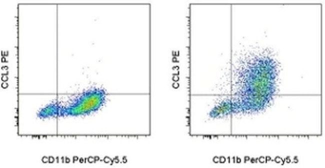
FIGURE: 1 / 2
CCL3 (MIP-1 alpha) Antibody (12-7532-82) in Flow


Product Details
12-7532-82
Species Reactivity
Published species
Host/Isotype
Recommended Isotype Control
Class
Type
Clone
Conjugate
Excitation/Emission Max
Form
Concentration
Purification
Storage buffer
Contains
Storage conditions
Shipping conditions
RRID
Product Specific Information
Description: This DNT3CC monoclonal antibody reacts with mouse CCL3. CCL3, also known as MIP-1 alpha (Macrophage Inflammatory Protein 1 alpha), is a member of the CC- subfamily of chemokines and is most closely related to CCL4, or MIP-1 beta. These proteins play critical roles in the recruitment of leukocytes to the site of inflammation. While both CCL3 and CCL4 will attract monocytes, macrophages, and dendritic cells, CCL3 preferentially attracts CD8+ T cells, while CD4+ T cells are more responsive to CCL4. In addition to its chemotactic functions, CCL3 induces inflammatory cytokine secretion, mast cell degranulation, and NK cell activation. It has also been reported to inhibit hematopoetic stem cell proliferation and may be responsible for the maintenance of these cells in a quiescent state. CCL3 signaling is mediated by the G protein-coupled receptors CCR1, CCR4, and CCR5, which are shared with CCL4 and CCL5 (RANTES).
Applications Reported: This DNT3CC antibody has been reported for use in intracellular staining followed by flow cytometric analysis.
Applications Tested: This DNT3CC antibody has been tested by intracellular staining and flow cytometric analysis of mouse thioglycolate-elicited peritoneal macrophages using the Intracellular Fixation & Permeabilization Buffer Set (Product # 88-8824-00) and protocol. Please refer to Best Protocols: Protocol A: Two step protocol for (cytoplasmic) intracellular proteins. This can be used at less than or equal to 0.125 µg per test. A test is defined as the amount (µg) of antibody that will stain a cell sample in a final volume of 100 µL. Cell number should be determined empirically but can range from 10^5 to 10^8 cells/test. It is recommended that the antibody be carefully titrated for optimal performance in the assay of interest.
Excitation: 488-561 nm; Emission: 578 nm; Laser: Blue Laser, Green Laser, Yellow-Green Laser.
Filtration: 0.2 µm post-manufacturing filtered.
Target Information
Both MIP-1alpha and MIP-1beta are structurally and functionally related CC chemokines. They participate in host response to invading bacterial, viral, parasite and fungal pathogens by regulating the trafficking and activation state of selected subgroups of inflammatory cells (e.g. macrophages, lymphocytes and NK cells). While both MIP-1alpha and MIP-1beta exert similar effects on monocytes, their effect on lymphocytes differ; with MIP-1alpha selectively attracting CD8+ lymphocytes, and MIP-1beta selectively attracting CD4+ lymphocytes. Additionally, MIP-1alpha and MIP-1beta have also been shown to be potent chemoattractants for B cells, eosinophils and dendritic cells. Both human and murine MIP-1alpha and MIP-1beta are active on human and murine hematopoietic cells.
For Research Use Only. Not for use in diagnostic procedures. Not for resale without express authorization.
How to use the Panel Builder
Watch the video to learn how to use the Invitrogen Flow Cytometry Panel Builder to build your next flow cytometry panel in 5 easy steps.
Bioinformatics
Protein Aliases: C-C motif chemokine 3; H-MIP-1-alpha; Heparin-binding chemotaxis protein; L2G25B; LD78-alpha; Macrophage inflammatory protein 1-alpha; macrophage inflammatory protein-1alpha; MIP-1 alpha; MIP-1-alpha; MIP1 (a); mip1 alpha; RP23-320E6.7; SIS-alpha; small inducible cytokine A3; Small-inducible cytokine A3; TY-5
Gene Aliases: AI323804; Ccl3; G0S19-1; LD78alpha; MIP-1alpha; MIP1-(a); MIP1-alpha; Mip1a; Scya3
UniProt ID: (Mouse) P10855
Entrez Gene ID: (Mouse) 20302

Performance Guarantee
If an Invitrogen™ antibody doesn't perform as described on our website or datasheet,we'll replace the product at no cost to you, or provide you with a credit for a future purchase.*
Learn more
We're here to help
Get expert recommendations for common problems or connect directly with an on staff expert for technical assistance related to applications, equipment and general product use.
Contact tech support

