Search Thermo Fisher Scientific
Invitrogen
Granzyme A Monoclonal Antibody (GzA-3G8.5), PE-Cyanine7, eBioscience™
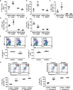
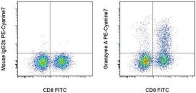
FIGURE: 1 / 2
Granzyme A Antibody (25-5831-82) in Flow


Product Details
25-5831-82
Species Reactivity
Published species
Host/Isotype
Recommended Isotype Control
Class
Type
Clone
Conjugate
Excitation/Emission Max
Form
Concentration
Purification
Storage buffer
Contains
Storage conditions
Shipping conditions
RRID
Product Specific Information
Description: This Gza-3G8.5 monoclonal antibody reacts with mouse Granzyme A. Granzyme A is the most abundantly expressed of the ten granzyme serine proteases that have been identified in mice. Granzymes are proteins released from the granules of NK cells and cytotoxic T lymphocytes that induce death in target cells by cleavage of intracellular substrates and play a critical role in immune defense against viruses, tumors, and intracellular bacteria. Granzyme A activates caspase-independent cell death pathways morphologically similar to apoptosis and characterized by mitochondrial and DNA damage. It may also play a role in inflammation, as the precursor form of IL-1 beta (pro-IL-1 beta) is among its target substrates. Granzyme A shares overlapping substrate specificity with the closely-related Granzyme K, which is believed to account for the minimal decrease in cytotoxicity of Granzyme A-deficient CTLs.
Applications Reported: This GzA-3G8.5 antibody has been reported for use in intracellular staining followed by flow cytometric analysis.
Applications Tested: This GzA-3G8.5 antibody has been tested by intracellular staining followed by flow cytometric analysis of mouse splenocytes. This can be used at less than or equal to 0.06 µg per test. A test is defined as the amount (µg) of antibody that will stain a cell sample in a final volume of 100 µL. Cell number should be determined empirically but can range from 10^5 to 10^8 cells/test. It is recommended that the antibody be carefully titrated for optimal performance in the assay of interest.
Light sensitivity: This tandem dye is sensitive to photo-induced oxidation. Please protect this vial and stained samples from light.
Fixation: Samples can be stored in IC Fixation Buffer (Product # 00-822-49) (100 µL of cell sample + 100 µL of IC Fixation Buffer) or 1-step Fix/Lyse Solution (Product # 00-5333-54) for up to 3 days in the dark at 4°C with minimal impact on brightness and FRET efficiency/compensation. Some generalizations regarding fluorophore performance after fixation can be made, but clone specific performance should be determined empirically.
Excitation: 488-561 nm; Emission: 775 nm; Laser: Blue Laser, Green Laser, Yellow-Green Laser.
Filtration: 0.2 µm post-manufacturing filtered.
Target Information
Cytolytic T lymphocytes (CTL) and natural killer (NK) cells share the remarkable ability to recognize, bind, and lyse specific target cells. They are thought to protect their host by lysing cells bearing on their surface 'nonself' antigens, usually peptides or proteins resulting from infection by intracellular pathogens. The protein described here is a T cell- and natural killer cell-specific serine protease that may function as a common component necessary for lysis of target cells by cytotoxic T lymphocytes and natural killer cells.
For Research Use Only. Not for use in diagnostic procedures. Not for resale without express authorization.
How to use the Panel Builder
Watch the video to learn how to use the Invitrogen Flow Cytometry Panel Builder to build your next flow cytometry panel in 5 easy steps.
Bioinformatics
Protein Aliases: Autocrine thymic lymphoma granzyme-like serine protease; BLT esterase; CTLA-3; Fragmentin-1; Granzyme A; H factor; Hanukah factor; serine esterase 1; T cell-specific serine protease 1; TSP-1
Gene Aliases: AW494114; Ctla-3; Ctla3; Gzma; Hf; Mtsp-1; SE1; TSP-1; TSP1
UniProt ID: (Mouse) P11032
Entrez Gene ID: (Mouse) 14938

Performance Guarantee
If an Invitrogen™ antibody doesn't perform as described on our website or datasheet,we'll replace the product at no cost to you, or provide you with a credit for a future purchase.*
Learn more
We're here to help
Get expert recommendations for common problems or connect directly with an on staff expert for technical assistance related to applications, equipment and general product use.
Contact tech support

