Search Thermo Fisher Scientific
Invitrogen
IL-1 beta Monoclonal Antibody (CRM56), Brilliant Violet™ 650, eBioscience™
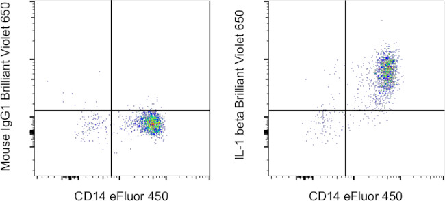
FIGURE: 1 / 1
IL-1 beta Antibody (416-7018-42) in Flow

Product Details
416-7018-42
Species Reactivity
Host/Isotype
Recommended Isotype Control
Class
Type
Clone
Conjugate
Excitation/Emission Max
Form
Concentration
Purification
Storage buffer
Contains
Storage conditions
Shipping conditions
RRID
Product Specific Information
Description: The CRM56 antibody reacts with human and baboon interleukin-1 beta; (IL-1 beta). IL-1 beta is a 17 kDa factor secreted primarily by monocytes. IL-1 has effects on T-helper cells, inducing IL-2 secretion and expression of IL-2 receptors. IL-1 has effects on B cells, promoting cell proliferation and immunoglobulin synthesis.
Applications Reported: This CRM56 antibody has been reported for use in intracellular staining followed by flow cytometric analysis.
Applications Tested: This CRM56 antibody has been pre-diluted and tested by intracellular staining followed by flow cytometric analysis of normal human peripheral blood cells using the Intracellular Fixation & Permeabilization Buffer Set (Product # 88-8824-00) and protocol. Please refer to "Staining Intracellular Antigens for Flow Cytometry, Protocol A: Two step protocol for intracellular (cytoplasmic) proteins" located at Flow Protocols. This may be used at 5 µL (0.25 µg) per test. A test is defined as the amount (µg) of antibody that will stain a cell sample in a final volume of 100 µL. Cell number should be determined empirically but can range from 10^5 to 10^8 cells/test.
Brilliant Violet™ 650 (BV650) is a tandem dye that emits at 649 nm and is intended for use on cytometers equipped with a violet (405 nm) laser. Please make sure that your instrument is capable of detecting this fluorochrome.
When using two or more Super Bright, Brilliant Violet™, Brilliant Ultra Violet™, or other polymer dye-conjugated antibodies in a staining panel, it is recommended to use Super Bright Complete Staining Buffer (Product # SB-4401-42) or Brilliant Stain Buffer™ (Product # 00-4409-75) to minimize any non-specific polymer interactions. Please refer to the datasheet for Super Bright Staining Buffer or Brilliant Stain Buffer for more information.
Light sensitivity: This tandem dye is sensitive to photo-induced oxidation. Please protect this vial and stained samples from light.
Fixation: Samples can be stored in IC Fixation Buffer (Product # 00-8222-49) (100 µL of cell sample + 100 µL of IC Fixation Buffer) or 1-step Fix/Lyse Solution (Product # 00-5333-54) for up to 3 days in the dark at 4°C with minimal impact on brightness and FRET efficiency/compensation. Some generalizations regarding fluorophore performance after fixation can be made, but clone-specific performance should be determined empirically.
Our internal testing suggests that Brilliant Violet™ 650 (BV650) is not compatible with methanol-based fixation.
Excitation: 407 nm; Emission: 649 nm; Laser: Violet Laser.
BRILLIANT VIOLET™ is a trademark or registered trademark of Becton, Dickinson and Company or its affiliates, and is used under license. Powered by Sirigen™.
Target Information
Interleukin-1 beta (IL-1 beta) is a proinflammatory cytokine expressed by monocytes, macrophages, and dendritic cells. IL-1 beta is synthesized in response to inflammatory stimuli as a 31 kDa inactive pro-form that accumulates in the cytosol. Cleavage of pro-IL-1 beta into the active 17 kDa protein requires the activation of inflammasomes, which are multi-protein complexes that respond to pathogens, stress conditions, and other danger signals. Inflammasome activation triggers the processing of the caspase-1 precursor into its active form, which in turn cleaves pro-IL-1 beta. IL-1 beta lacks a signal sequence peptide for classical ER/Golgi pathway and is secreted alongside caspase-1 via an alternate and incompletely understood mechanism. Although IL-1 beta is most often secreted in its active form, secretion of the uncleaved protein may be detectable under some biological conditions. IL-1 beta signals through two receptors, IL-1RI and IL-1RII, both of which are shared with IL-1 alpha. IL-1 beta activity can be moderated by IL-1 Receptor Antagonist (IL-1RA), a protein produced by many cell types that blocks receptor binding through competitive inhibition. IL-1 beta play an important role in innate host defense by triggering the production of other proinflammatory cytokines in target cells and initiating acute-phase responses to infection and injury. Elevated levels of IL-1 beta have been associated with many chronic inflammatory conditions IL-1 beta neutralizing antibodies potential therapeutic value.
For Research Use Only. Not for use in diagnostic procedures. Not for resale without express authorization.
How to use the Panel Builder
Watch the video to learn how to use the Invitrogen Flow Cytometry Panel Builder to build your next flow cytometry panel in 5 easy steps.
References (0)
Bioinformatics
Protein Aliases: Catabolin; Hematopoietin 1 (H1); IFN beta inducing factor; il 1b; IL 1β; IL-1 beta; IL1 B; IL1β; ILN; Interleukin; Interleukin 1 beta precursor; interleukin 1, beta; Interleukin-1 beta; Interleukin-1b; Interleukin1 beta; LAF; Osteoclast activating factor (OAF); preinterleukin 1 beta; Pro interleukin 1 beta; pro-interleukin-1-beta
Gene Aliases: IL-1; IL1-BETA; IL1B; IL1F2
UniProt ID: (Human) P01584
Entrez Gene ID: (Human) 3553

Performance Guarantee
If an Invitrogen™ antibody doesn't perform as described on our website or datasheet,we'll replace the product at no cost to you, or provide you with a credit for a future purchase.*
Learn more
We're here to help
Get expert recommendations for common problems or connect directly with an on staff expert for technical assistance related to applications, equipment and general product use.
Contact tech support

