Search Thermo Fisher Scientific
Invitrogen
GFP Monoclonal Antibody (5F12.4), eFluor™ 660, eBioscience™
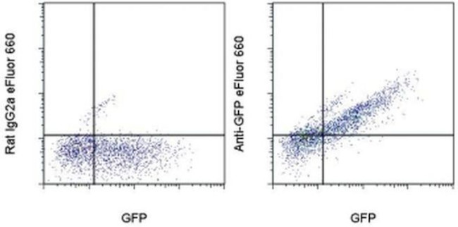
FIGURE: 1 / 1
GFP Antibody (50-6498-82) in Flow

Product Details
50-6498-82
Species Reactivity
Published species
Host/Isotype
Recommended Isotype Control
Class
Type
Clone
Conjugate
Excitation/Emission Max
Form
Concentration
Purification
Storage buffer
Contains
Storage conditions
Shipping conditions
RRID
Product Specific Information
Description: This 5F12.4 monoclonal antibody reacts with green-fluorescent protein (GFP), which was originally isolated from the cnidarian Aequorea victoria. This protein absorbs blue light (maximally at 395 nm) and emits green light (peak at 509) without the requirement of exogenous substrates and cofactors. These unique qualities allow GFP to be used to monitor gene expression and protein localization in vivo. Several mutant forms of GFP have been developed which fluoresce more intensely and have shifted excitation maxima when compared to wildtype GFP, making them useful for flow cytometry, fluorescence microscopy, and double-labeling applications. This antibody is capable of staining formaldehyde fixed cells.
Applications Reported: This 5F12.4 antibody has been reported for use in flow cytometric analysis.
Applications Tested: This 5F12.4 antibody has been tested by flow cytometric analysis of GFP expressing cells. This can be used at less than or equal to 0.125 µg per test. A test is defined as the amount (µg) of antibody that will stain a cell sample in a final volume of 100 µL. Cell number should be determined empirically but can range from 10^5 to 10^8 cells/test. It is recommended that the antibody be carefully titrated for optimal performance in the assay of interest.
eFluor® 660 is a replacement for Alexa Fluor® 647. eFluor® 660 emits at 659 nm and is excited with the red laser (633 nm). Please make sure that your instrument is capable of detecting this fluorochome.
Excitation: 633-647 nm; Emission: 668 nm; Laser: Red Laser.
Filtration: 0.2 µm post-manufacturing filtered.
Target Information
The jellyfish Aequorea victoria contains green fluorescent protein (GFP Tag) that emits light in the bioluminescence reaction of the animal. GFP has been used widely as a reporter protein for gene expression in eukaryotic and prokaryotic organisms, and as a protein tag in cell culture and in multicellular organisms. As a fusion tag, GFP can be used to localize proteins, to study their movement or to research the dynamics of the subcellular compartments where these proteins are targeted. GFP technology has revealed considerable new insights in the physiological activities of living cells. GFP is a 27 kDa monomeric protein, which autocatalytically forms a fluorescent pigment. The wild type protein absorbs blue light (maximally at 395nm) and emits green light (peak emission 508nm) in the absence of additional proteins, substrates, or co-factors. GFP fluorescence is stable, species independent and is suitable for a variety of applications. GFP has been used extensively as a fluorescent tag to monitor gene expressin and protein localization. Moreover, other applications for GFP include its use in assessing protein protein interactions in the yeast two hybrid system, and in measuring distances between proteins in fluorescence energy transfer (FRET) experiments.
For Research Use Only. Not for use in diagnostic procedures. Not for resale without express authorization.
How to use the Panel Builder
Watch the video to learn how to use the Invitrogen Flow Cytometry Panel Builder to build your next flow cytometry panel in 5 easy steps.
Bioinformatics
Protein Aliases: GFP; GFP tag; GFP2; green fluorescence; green fluorescent; Turbo eGFP

Performance Guarantee
If an Invitrogen™ antibody doesn't perform as described on our website or datasheet,we'll replace the product at no cost to you, or provide you with a credit for a future purchase.*
Learn more
We're here to help
Get expert recommendations for common problems or connect directly with an on staff expert for technical assistance related to applications, equipment and general product use.
Contact tech support

