Search Thermo Fisher Scientific
Invitrogen
CD183 (CXCR3) Monoclonal Antibody (CXCR3-173), Super Bright™ 600, eBioscience™
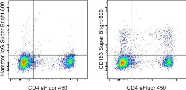
FIGURE: 1 / 13
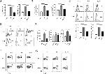
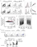
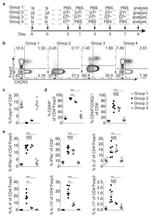
CD183 (CXCR3) Antibody (63-1831-82) in Flow







Product Details
63-1831-82
Species Reactivity
Published species
Host/Isotype
Recommended Isotype Control
Class
Type
Clone
Conjugate
Excitation/Emission Max
Form
Concentration
Purification
Storage buffer
Contains
Storage conditions
Shipping conditions
RRID
Product Specific Information
Description: The monoclonal antibody CXCR3-173 recognizes mouse CD183 also known as CXCR3. CD183 is a seven transmembrane G-protein liked chemokine receptor which binds three ligands; CXCL9 (mig), CXCL10 (IP-10)and CXCL11 (ITAC). CD183 as been shown to play a role in CD4 T cell responses to grafts. CXCR3 knockout mice have compromised allograft rejection responses. Expression is found on NK cells, a subset of T lymphocytes and a subset of Tregs as well as preferential expression on Th1-polarized cells.
The antibody CXCR3-173 has been shown to affect chemotaxis in response to ligand. The presence of ligand eliminates staining with the antibody. In vivo addition of the antibody delays cardiac and pancreatic islet allograft rejection.
Applications Reported: This CXCR3-173 antibody has been reported for use in flow cytometric analysis.
Applications Tested: This CXCR3-173 antibody has been tested by flow cytometric analysis of mouse splenocytes. This can be used at less than or equal to 1.0 µg per test. A test is defined as the amount (µg) of antibody that will stain a cell sample in a final volume of 100 µL. Cell number should be determined empirically but can range from 10^5 to 10^8 cells/test. It is recommended that the antibody be carefully titrated for optimal performance in the assay of interest.
Super Bright 600 is a tandem dye that can be excited with the violet laser line (405 nm) and emits at 600 nm. We recommend using a 610/20 bandpass filter. Please make sure that your instrument is capable of detecting this fluorochrome.
When using two or more Super Bright dye-conjugated antibodies in a staining panel, it is recommended to use Super Bright Complete Staining Buffer (Product # SB-4401) to minimize any non-specific polymer interactions. Please refer to the datasheet for Super Bright Staining Buffer for more information.
Light sensitivity: This tandem dye is sensitive to photo-induced oxidation. Please protect this vial and stained samples from light.
Fixation: Samples can be stored in IC Fixation Buffer (Product # 00-8222) (100 µL of cell sample + 100 µL of IC Fixation Buffer) or 1-step Fix/Lyse Solution (Product # 00-5333) for up to 3 days in the dark at 4°C with minimal impact on brightness and FRET efficiency/compensation. Some generalizations regarding fluorophore performance after fixation can be made, but clone specific performance should be determined empirically.
Excitation: 405 nm; Emission: 600 nm; Laser: Violet Laser
Super Bright Polymer Dyes are sold under license from Becton, Dickinson and Company.
Target Information
This gene encodes a G protein-coupled receptor with selectivity for three chemokines, termed IP10 (interferon-g-inducible 10 kDa protein), Mig (monokine induced by interferon-g) and I-TAC (interferon-inducible T cell a-chemoattractant). IP10, Mig and I-TAC belong to the structural subfamily of CXC chemokines, in which a single amino acid residue separates the first two of four highly conserved Cys residues. Binding of chemokines to this protein induces cellular responses that are involved in leukocyte traffic, most notably integrin activation, cytoskeletal changes and chemotactic migration. Inhibition by Bordetella pertussis toxin suggests that heterotrimeric G protein of the Gi-subclass couple to this protein. Signal transduction has not been further analyzed but may include the same enzymes that were identified in the signaling cascade induced by other chemokine receptors. As a consequence of chemokine-induced cellular desensitization (phosphorylation-dependent receptor internalization), cellular responses are typically rapid and short in duration. Cellular responsiveness is restored after dephosphorylation of intracellular receptors and subsequent recycling to the cell surface. This gene is prominently expressed in in vitro cultured effector/memory T cells, and in T cells present in many types of inflamed tissues. In addition, IP10, Mig and I-TAC are commonly produced by local cells in inflammatory lesion, suggesting that this gene and its chemokines participate in the recruitment of inflammatory cells. Therefore, this protein is a target for the development of small molecular weight antagonists, which may be used in the treatment of diverse inflammatory diseases. Multiple transcript variants encoding different isoforms have been found for this gene.
For Research Use Only. Not for use in diagnostic procedures. Not for resale without express authorization.
How to use the Panel Builder
Watch the video to learn how to use the Invitrogen Flow Cytometry Panel Builder to build your next flow cytometry panel in 5 easy steps.
Bioinformatics
Protein Aliases: an; C Cmotif chemokine; C X C motif chemokine; C-X-C chemokine receptor type 3; CC motif chemokine; CCmotif chemokine; CD183; CXC; CXC motif chemokine; CXC-R3; Interferon-inducible protein 10 receptor; IP-10 receptor; IP10; IP10 receptor
Gene Aliases: Cd183; Cmkar3; Cxcr3
UniProt ID: (Mouse) O88410
Entrez Gene ID: (Mouse) 12766

Performance Guarantee
If an Invitrogen™ antibody doesn't perform as described on our website or datasheet,we'll replace the product at no cost to you, or provide you with a credit for a future purchase.*
Learn more
We're here to help
Get expert recommendations for common problems or connect directly with an on staff expert for technical assistance related to applications, equipment and general product use.
Contact tech support
