Search Thermo Fisher Scientific
Invitrogen
CD49b (Integrin alpha 2) Monoclonal Antibody (P1H5), APC, eBioscience™
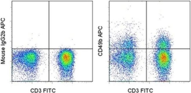
FIGURE: 1 / 1
CD49b (Integrin alpha 2) Antibody (17-0500-42) in Flow

Product Details
17-0500-42
Species Reactivity
Published species
Host/Isotype
Recommended Isotype Control
Class
Type
Clone
Conjugate
Excitation/Emission Max
Form
Concentration
Purification
Storage buffer
Contains
Storage conditions
Shipping conditions
RRID
Product Specific Information
Description: This P1H5 monoclonal antibody reacts with human CD49b, which is also known as integrin alpha 2. Integrins constitute a family of cell surface receptors that mediate adhesion between cells or between a cell and the extracellular matrix. These proteins are heterodimers consisting of two distinct subunits, alpha and beta. Integrins are expressed on hematopoietic, non-hematopoietic, and cancer cells. Integrin-mediated adhesion is regulated by changes in ligand affinity (or activation), cell shape, and integrin clustering and diffusion. Integrin alpha 2 forms a complex with the common beta 1 subunit to form the alpha2beta1 integrin, which binds type I collagen and laminin. Moreover, studies suggest that this complex suppresses metastasis in cancer and participates in T cell costimulation by activating the MAPK and PI3K pathways.
The P1H5 monoclonal antibody has been reported to inhibit adhesion of fibroblasts and platelets to collagen. Crossblocking studies indicate that P1H5 and eBioY418 recognize different epitopes.
Applications Reported: This P1H5 antibody has been reported for use in flow cytometric analysis.
Applications Tested: This P1H5 antibody has been pre-titrated and tested by flow cytometric analysis of normal human peripheral blood cells. This can be used at 5 µL (0.125 µg) per test. A test is defined as the amount (µg) of antibody that will stain a cell sample in a final volume of 100 µL. Cell number should be determined empirically but can range from 10^5 to 10^8 cells/test.
Excitation: 633-647 nm; Emission: 660 nm; Laser: Red Laser.
Filtration: 0.2 µm post-manufacturing filtered.
Target Information
CD49b (Integrin alpha 2, Integrin alpha 2 chain, VLA-2 alpha chain, HM alpha 2) is a member of the integrin family. It is a glycoprotein with molecular weight of 150 kD, and it complexes with CD29 (Integrin beta 1) to form the heterodimeric integrin VLA-2 (integrin alpha 2 beta 1, or GPIa/IIa) complex. VLA-2 is an extracellular receptor for laminin, collagen, and fibronectin, and interaction with its ligands results in the activation of intracellular signaling pathways. It has reported roles in VEGF-induced angiogenesis in vivo, as well as adhesion and lymphocyte activation. CD49b is expressed by NK cells, NK-T cells, monocytes, platelets, and epithelial cells. It is also expressed on adaptive immune cells such as T and B cells, specifically on a subset of CD4+ T cells in the spleen, on intraepithelial and lamina propria lymphocytes in the intestine, as well as on a population of peripheral CD4+ type 1 T regulatory (Tr1) cells that co-express LAG-3.
For Research Use Only. Not for use in diagnostic procedures. Not for resale without express authorization.
How to use the Panel Builder
Watch the video to learn how to use the Invitrogen Flow Cytometry Panel Builder to build your next flow cytometry panel in 5 easy steps.
Bioinformatics
Protein Aliases: alpha 2 subunit of VLA-2 receptor; BDPLT9; CD49 antigen-like family member B; CD49b; Collagen receptor; GPIa; human platelet alloantigen system 5; Integrin alpha-2; integrin, alpha 2 (CD49B, alpha 2 subunit of VLA-2 receptor); platelet antigen Br; platelet glycoprotein GPIa; Platelet membrane glycoprotein Ia; very late activation protein 2 receptor, alpha-2 subunit; VLA-2 subunit alpha; VLA2 receptor
Gene Aliases: BR; CD49B; GPIa; HPA-5; ITGA2; VLA-2; VLAA2
UniProt ID: (Human) P17301
Entrez Gene ID: (Human) 3673

Performance Guarantee
If an Invitrogen™ antibody doesn't perform as described on our website or datasheet,we'll replace the product at no cost to you, or provide you with a credit for a future purchase.*
Learn more
We're here to help
Get expert recommendations for common problems or connect directly with an on staff expert for technical assistance related to applications, equipment and general product use.
Contact tech support

