Search Thermo Fisher Scientific
Invitrogen
EGFR Monoclonal Antibody (me1B3), eFluor™ 660, eBioscience™
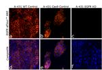
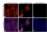
This Antibody was verified by Knockout to ensure that the antibody binds to the antigen stated.
FIGURE: 1 / 3
EGFR Antibody (50-9509-42) in ICC/IF


Product Details
50-9509-42
Species Reactivity
Host/Isotype
Recommended Isotype Control
Class
Type
Clone
Conjugate
Excitation/Emission Max
Form
Concentration
Purification
Storage buffer
Contains
Storage conditions
Shipping conditions
RRID
Product Specific Information
Description: This me1B3 monoclonal antibody reacts with human epidermal growth factor receptor (EGFR), also known as ErbB1 and Her1. EGFR is a receptor tyrosine kinase belonging to the EGFR/ErbB receptor subfamily, which also includes ErbB2/Her2/neu. EGFR contains an extracellular ligand-binding domain, a single transmembrane-spanning region, and a cytoplasmic tail containing a tyrosine kinase domain. Specific ligands include EGF and TNF alpha, as well as other members of the EGF protein family. Upon ligand binding, EGFR undergoes receptor-mediated dimerization with itself or ErbB2/Her2/neu. Dimerization induces auto-phosphorylation, resulting in the activation of PLC gamma, JAK/STAT, MAPK/ERK, and PI3K/AKT signaling pathways. EGFR has a role in cell proliferation, migration and differentiation, and is evolutionarily highly conserved in structure and function. EGFR is broadly expressed and is overexpressed or mutated in glioblastomas and a number of different epithelial cancers, including non-small cell lung cancer and colorectal cancer.
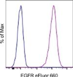
Applications Reported: This me1B3 antibody has been reported for use in flow cytometric analysis.
Applications Tested: This me1B3 antibody has been pre-titrated and tested by flow cytometric analysis of HeLa cells. This can be used at 5 µL (0.125 µg) per test. A test is defined as the amount (µg) of antibody that will stain a cell sample in a final volume of 100 µL. Cell number should be determined empirically but can range from 10^5 to 10^8 cells/test.
eFluor® 660 is a replacement for Alexa Fluor® 647. eFluor® 660 emits at 659 nm and is excited with the red laser (633 nm). Please make sure that your instrument is capable of detecting this fluorochome.
Excitation: 633-647 nm; Emission: 668 nm; Laser: Red Laser.
Filtration: 0.2 µm post-manufacturing filtered.
Target Information
EGFR, epidermal growth factor receptor, is a receptor tyrosine kinases that signals in response to various growth factors. Overexpression has been linked to numerous types of cancer and EGFR is the target of both biological and small molecular therapeutics. EGFR is encoded by the EGFR gene located on chromosome 7 in humans. EGFR belongs to the HER/ERbB family of proteins that includes three other receptor tyrosine kinases, ERbB2, ERbB3, ERbB4. EGFR is a transmembrane receptor and binding of its cognate ligands such as EGF (Epidermal Growth Factor) and TGF alpha (Transforming Growth Factor alpha) to the extracellular domain leads to EGFR dimerization followed by autophosphorylation of the tyrosine residues in the cytoplasmic domain. Overexpression is observed in tumors of the head and neck, brain, bladder, stomach, breast, lung, endometrium, cervix, vulva, ovary, esophagus, stomach and in squamous cell carcinoma.
For Research Use Only. Not for use in diagnostic procedures. Not for resale without express authorization.
How to use the Panel Builder
Watch the video to learn how to use the Invitrogen Flow Cytometry Panel Builder to build your next flow cytometry panel in 5 easy steps.
References (0)
Bioinformatics
Protein Aliases: 2.7.10.1; avian erythroblastic leukemia viral (v-erb-b) oncogene homolog; cell growth inhibiting protein 40; cell proliferation-inducing protein 61; EC 2.7.10.1; Epidermal growth factor receptor; erb-b2 receptor tyrosine kinase 1; kinase EGFR; Oncogene ERBB; Proto-oncogene c-ErbB-1; Receptor tyrosine-protein kinase erbB-1; Urogastrone
Gene Aliases: EGFR; ERBB; ERBB1; HER1; mENA; NISBD2; PIG61
UniProt ID: (Human) P00533
Entrez Gene ID: (Human) 1956

Performance Guarantee
If an Invitrogen™ antibody doesn't perform as described on our website or datasheet,we'll replace the product at no cost to you, or provide you with a credit for a future purchase.*
Learn more
We're here to help
Get expert recommendations for common problems or connect directly with an on staff expert for technical assistance related to applications, equipment and general product use.
Contact tech support

