Search Thermo Fisher Scientific
Invitrogen
GATA-3 Recombinant Rat Monoclonal Antibody (TWAJ), Alexa Fluor™ Plus 555
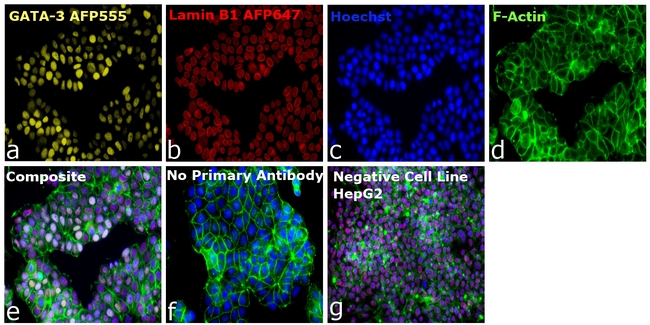
This Antibody was verified by Relative expression to ensure that the antibody binds to the antigen stated.
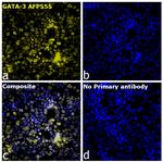
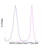
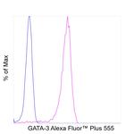
FIGURE: 1 / 6
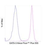
GATA-3 Antibody (740023TP555) in ICC/IF


Product Details
740023TP555
Species Reactivity
Host/Isotype
Expression System
Class
Type
Clone
Conjugate
Excitation/Emission Max
Form
Concentration
Purification
Storage buffer
Contains
Storage conditions
Shipping conditions
Product Specific Information
Alexa Fluor™ Plus recombinant antibodies are conjugated using new, proprietary dye chemistry so you can generate stunning data. Alexa Fluor™ Plus antibodies represent an advancement in fluorescent conjugate technology. Alexa Fluor™ Plus antibodies provide brighter signal compared to leading Alexa Fluor™ antibodies, providing you with better signal-to-noise for your critical experiments. These antibodies show better specificity and lot-to-lot consistency as these are recombinant antibodies, generated by cloning specific genes for the desired antibodies into an expression vector and expressed in vitro.
Using conjugate solutions: Centrifuge the protein conjugate solution briefly in a microcentrifuge before use; add only the supernatant to the experiment. This step will help eliminate any protein aggregates that may have formed during storage, thereby reducing nonspecific background staining.
Applications Tested: This TWAJ antibody has been tested by immunocytochemistry and flow cytometric analysis of MCF-7 cells. This may be used for immunocytochemistry at 2 µg/ml and for flow cytometry at less than or equal to 0.5 µg per test. A test is defined as the amount (µg) of antibody that will stain a cell sample in a final volume of 100 µL. Cell number should be determined empirically but can range from 10^5 to 10^8 cells/test. It is recommended that the antibody be carefully titrated for optimal performance in the assay of interest.
Excitation: 553 nm; Emission: 568 nm; Laser: Yellow Laser
Filtration: 0.2 µm post-manufacturing filtered.
Target Information
The genes for all 4 subunits of the T-cell antigen receptor (alpha, beta, gamma and delta) are controlled by distinct enhancers and their enhancer-binding proteins. Marine and Winoto (1991) identified a common TCR regulatory element by demonstrating binding of the enhancer-binding protein GATA3 to the enhancer elements of all 4 TCR genes. GATA3 had been shown in the chicken to be an enhancer-binding protein containing a zinc finger domain. GATA3 mRNA was demonstrated by Northern blot analysis in T cells but not in B cells or macrophages. GATA3 is abundantly expressed in the T-lymphocyte lineage and is thought to participate in T-cell receptor gene activation through binding to enhancers. Labastie et al. (1994) cloned the human gene and the 5-prime end of the mouse gene. The human gene comprises 6 exons distributed over 17 kb of DNA. Its 2 zinc fingers are encoded by 2 separate exons highly conserved with those of GATA1, but no other structural homologies between the 2 genes could be found.
For Research Use Only. Not for use in diagnostic procedures. Not for resale without express authorization.
References (0)
Bioinformatics
Protein Aliases: GATA binding protein-3; GATA-binding factor 3; MGC2346; MGC5199; MGC5445; Trans-acting T-cell-specific transcription factor GATA-3; Transacting T-cell-specific transcription factor GATA-3
Gene Aliases: GATA3; HDR; HDRS
UniProt ID: (Human) P23771
Entrez Gene ID: (Human) 2625

Performance Guarantee
If an Invitrogen™ antibody doesn't perform as described on our website or datasheet,we'll replace the product at no cost to you, or provide you with a credit for a future purchase.*
Learn more
We're here to help
Get expert recommendations for common problems or connect directly with an on staff expert for technical assistance related to applications, equipment and general product use.
Contact tech support
