Search Thermo Fisher Scientific
Invitrogen
GFP Polyclonal Antibody, Alexa Fluor™ 555
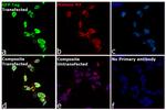
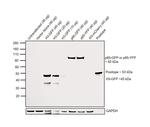
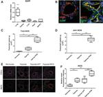
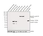
This Antibody was verified by Relative expression to ensure that the antibody binds to the antigen stated.
FIGURE: 1 / 12
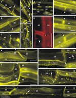
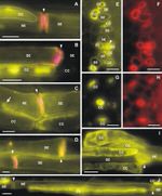
GFP Antibody (A-31851) in ICC/IF

Product Details
A-31851
Species Reactivity
Published species
Host/Isotype
Class
Type
Immunogen
Conjugate
Excitation/Emission Max
Form
Concentration
Purification
Storage buffer
Contains
Storage conditions
Shipping conditions
RRID
Product Specific Information
At the time of preparation, the products are certified to be free of unconjugated dyes and are tested in a cytological experiment to ensure low nonspecific staining. Several Alexa Fluor™ dye-conjugates made from the rabbit anti-GFP IgG fraction are also available. The Alexa Fluor™ dyes provide for extraordinarily bright antibody conjugates. The approximate fluorescence excitation (Ex) and emission maxima for this Alexa Fluor™ 555 conjugate are 555 nm and 565 nm, respectively.
These anti-GFP antibody conjugates are suited for detection of native GFP, GFP variants, and most GFP-fusion proteins by western blot analysis and immunocytochemistry. For initial experiments, we recommend trying dilutions ranging from 1:200 to 1:2,000 with our fluorophore-labeled antibodies, a final concentration of 1-10 µg/mL should be satisfactory for most immunocytochemical applications. If using the rabbit anti-GFP antibody-Alexa Fluor™ conjugates in western blotting, dilute the conjugate 1:2,000 in PBS to obtain a final antibody concentration of 1 µg/mL. HeLa and U2-OS cells were tested with this antibody in immunocytochemistry, but not with paraffin-embedded sections.
It is a good practice to centrifuge the protein conjugate solutions briefly in a microcentrifuge before use; add only the supernatant to the experiment. This step eliminates any protein aggregates that may have formed during storage, thereby reducing nonspecific background staining.
Target Information
The jellyfish Aequorea victoria contains green fluorescent protein (GFP Tag) that emits light in the bioluminescence reaction of the animal. GFP has been used widely as a reporter protein for gene expression in eukaryotic and prokaryotic organisms, and as a protein tag in cell culture and in multicellular organisms. As a fusion tag, GFP can be used to localize proteins, to study their movement or to research the dynamics of the subcellular compartments where these proteins are targeted. GFP technology has revealed considerable new insights in the physiological activities of living cells. GFP is a 27 kDa monomeric protein, which autocatalytically forms a fluorescent pigment. The wild type protein absorbs blue light (maximally at 395nm) and emits green light (peak emission 508nm) in the absence of additional proteins, substrates, or co-factors. GFP fluorescence is stable, species independent and is suitable for a variety of applications. GFP has been used extensively as a fluorescent tag to monitor gene expressin and protein localization. Moreover, other applications for GFP include its use in assessing protein protein interactions in the yeast two hybrid system, and in measuring distances between proteins in fluorescence energy transfer (FRET) experiments.
For Research Use Only. Not for use in diagnostic procedures. Not for resale without express authorization.
Bioinformatics
Protein Aliases: GFP; GFP tag; GFP2; green fluorescence; green fluorescent; Turbo eGFP

Performance Guarantee
If an Invitrogen™ antibody doesn't perform as described on our website or datasheet,we'll replace the product at no cost to you, or provide you with a credit for a future purchase.*
Learn more
We're here to help
Get expert recommendations for common problems or connect directly with an on staff expert for technical assistance related to applications, equipment and general product use.
Contact tech support

