Search Thermo Fisher Scientific
Invitrogen
MAVS Monoclonal Antibody (ABM28H9), eFluor™ 450, eBioscience™
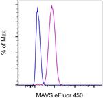
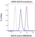
This Antibody was verified by Knockdown to ensure that the antibody binds to the antigen stated.
FIGURE: 1 / 2
MAVS Antibody (48-9835-42) in Flow


Product Details
48-9835-42
Species Reactivity
Host/Isotype
Recommended Isotype Control
Class
Type
Clone
Immunogen
Conjugate
Excitation/Emission Max
Form
Concentration
Purification
Storage buffer
Contains
Storage conditions
Shipping conditions
RRID
Product Specific Information
Description: This ABM28H9 monoclonal antibody recognizes human MAVS, a mitochondrial membrane protein also known as VISA, CARDIF or IPS-1.
Applications Reported: This ABM18H9 antibody has been reported for use in intracellular staining followed by flow cytometric analysis.
Applications Tested: This ABM18H9 antibody has been pre-diluted and tested by intracellular staining followed by flow cytometric analysis of HEL cells using the Intracellular Fixation & Permeabilization Buffer Set (Product # 88-8824-00) and protocol. Please refer to "Staining Intracellular Antigens for Flow Cytometry, Protocol A: Two step protocol for intracellular (cytoplasmic) proteins" located at www.thermofisher.com/flowprotocols . This may be used at 5 µL (0.125 µg) per test. A test is defined as the amount (µg) of antibody that will stain a cell sample in a final volume of 100 µL. Cell number should be determined empirically but can range from 10^5 to 10^8 cells/test.
Light sensitivity: This tandem dye is sensitive to photo-induced oxidation. Please protect this vial and stained samples from light.
Fixation: Samples can be stored in IC Fixation Buffer (Product # 00-8222-49) (100 µL of cell sample + 100 µL of IC Fixation Buffer) or 1-step Fix/Lyse Solution (Product # 00-5333-57) for up to 3 days in the dark at 4°C with minimal impact on brightness and FRET efficiency/compensation. Some generalizations regarding fluorophore performance after fixation can be made, but clone specific performance should be determined empirically.
Excitation: 488-561 nm; Emission: 775 nm; Laser: Blue Laser, Green Laser, Yellow-Green Laser.
Target Information
Two distinct signaling pathways activate the host innate immunity against viral infection. One pathway is reliant on members of the Toll-like receptor (TLR) family while the other uses the RNA helicase RIG-I as a receptor for intracellular viral double-stranded RNA as a trigger for the immune response. MAVS is a mitochondrial membrane protein that was identified as a critical component in the IFN beta signaling pathways that recruits IRF-3 to RIG-I, leading to its activation and that of NF-kappa-B. MAVS is also thought to interact with other components of the innate immune pathway such as the TLR adapter protein TRIF, TRAF2 and TRAF6. MAVS also interacts with the IKK-alpha, IKK-beta and IKK-iota kinases through its C-terminal region. Cleavage of this region by the Hepatitis C virus (HCV) protease allows HCV to escape the host immune system. Multiple isoforms of MAVS are known to exist.
For Research Use Only. Not for use in diagnostic procedures. Not for resale without express authorization.
How to use the Panel Builder
Watch the video to learn how to use the Invitrogen Flow Cytometry Panel Builder to build your next flow cytometry panel in 5 easy steps.
References (0)
Bioinformatics
Protein Aliases: CARD adapter inducing interferon beta; CARD adaptor inducing IFN-beta; Cardif; IFN-B promoter stimulator 1; Interferon beta promoter stimulator protein 1; IPS-1; MAVS; Mitochondrial antiviral-signaling protein; Putative NF-kappa-B-activating protein 031N; virus-induced signaling adaptor; Virus-induced-signaling adapter; VISA
Gene Aliases: CARDIF; IPS-1; IPS1; KIAA1271; MAVS; VISA
UniProt ID: (Human) Q7Z434
Entrez Gene ID: (Human) 57506

Performance Guarantee
If an Invitrogen™ antibody doesn't perform as described on our website or datasheet,we'll replace the product at no cost to you, or provide you with a credit for a future purchase.*
Learn more
We're here to help
Get expert recommendations for common problems or connect directly with an on staff expert for technical assistance related to applications, equipment and general product use.
Contact tech support

