Search Thermo Fisher Scientific
Invitrogen
Phospho-Histone H3 (Ser28) Monoclonal Antibody (HTA28), eFluor™ 660, eBioscience™
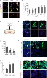
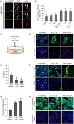
This Antibody was verified by Cell treatment to ensure that the antibody binds to the antigen stated.
FIGURE: 1 / 3
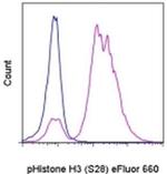
Phospho-Histone H3 (Ser28) Antibody (50-9124-42) in Flow


Product Details
50-9124-42
Species Reactivity
Published species
Host/Isotype
Recommended Isotype Control
Class
Type
Clone
Conjugate
Excitation/Emission Max
Form
Concentration
Purification
Storage buffer
Contains
Storage conditions
Shipping conditions
RRID
Product Specific Information
Description: The HTA28 monoclonal antibody recognizes phosphorylated serine 28 of human, mouse, rat, bovine, and hamster histone H3. This 15 kDa protein is a component of eukaryotic chromatin that is involved in forming the nucleosome structure. Histone H3 can be phosphorylated at serine 10 and serine 28. Studies have demonstrated that phosphorylation at serine 28 is mediated by MSK1 following activation by the MAP kinase signaling pathway in response to tumor promoters (e.g., UV and EGF) and oncoproteins (e.g., c-Myc, c-Jun, and c-Fos). Histone H3 serine 28 phosphorylation has been linked to chromosome condensation during mitosis, cell transformation, and regulation of RNA polymerase III transcription machinery.
Applications Reported: This HTA28 antibody has been reported for use in intracellular staining followed by flow cytometric analysis.
Applications Tested: This HTA28 antibody has been pre-titrated and tested by intracellular staining followed by flow cytometric analysis of nocodazole-treated HeLa cells. This can be used at 5 µL (0.25 µg) per test. A test is defined as the amount (µg) of antibody that will stain a cell sample in a final volume of 100 µL. Cell number should be determined empirically but can range from 10^5 to 10^8 cells/test.
Protocols: We recommend using Protocol A: Two-step protocol: intracellular (cytoplasmic) proteins or Protocol C: Two-step protocol: Fixation/Methanol. Protocol B: One-step protocol: intracellular (nuclear) proteins cannot be used. All Protocols can be found in the "Staining intracellular Antigens for Flow Cytometry Protocol" located in the Best Protocols Section under the Resources tab online.
eFluor® 660 is a replacement for Alexa Fluor® 647. eFluor® 660 emits at 659 nm and is excited with the red laser (633 nm). Please make sure that your instrument is capable of detecting this fluorochome.
Excitation: 633-647 nm; Emission: 668 nm; Laser: Red Laser.
Filtration: 0.2 µm post-manufacturing filtered.
Target Information
Histone H3 is one of the DNA-binding proteins found in the chromatin of all eukaryotic cells. H3 along with four core histone proteins binds to DNA forming the structure of the nucleosome. Histones play a central role in transcription regulation, DNA repair, DNA replication and chromosomal stability. Post translationally, histones are modified in a variety of ways to either directly change the chromatin structure or allow for the binding of specific transcription factors. The N-terminal tail of histone H3 protrudes from the globular nucleosome core and can undergo several different types of post-translational modification that influence cellular processes. These modifications include the covalent attachment of methyl or acetyl groups to lysine and arginine amino acids and the phosphorylation of serine or threonine.
For Research Use Only. Not for use in diagnostic procedures. Not for resale without express authorization.
How to use the Panel Builder
Watch the video to learn how to use the Invitrogen Flow Cytometry Panel Builder to build your next flow cytometry panel in 5 easy steps.
Bioinformatics
Protein Aliases: H3 histone family, member A; H3 histone family, member I; H3 histone family, member J; H3 histone family, member K; H3 histone family, member L; H3 histone family, member M; H3 histone, family 2; H3-clustered histone 13; H3-clustered histone 14; H3-clustered histone 15; H3-clustered histone 2; H3-clustered histone 3; H3-clustered histone 4; H3-clustered histone 6; H3-clustered histone 7; H3/b; H3/d; H3/f; H3/j; H3S10ph; histone 1, H3a; histone 1, H3b; histone 1, H3f; histone 1, H3h; histone 1, H3j; histone 2, H3a; histone 2, H3c; histone 2, H3c2; histone 2, H3ca2; Histone 3; Histone Cluster 2 H3a; histone cluster 2, H3c2, pseudogene; histone gene complex 1; histone H3; Histone H3.1; Histone H3.2; Histone H3/a; Histone H3/b; Histone H3/c; Histone H3/d; Histone H3/f; Histone H3/h; Histone H3/i; Histone H3/j; Histone H3/k; Histone H3/l; Histone H3/m; Histone H3/o
Gene Aliases: H3; H3-143; H3-291; H3-53; H3-614; H3-B; H3-F; H3.1-221; H3.1-291; H3.1-I; H3.2; H3.2-221; H3.2-614; H3.2-615; H3.2-616; H3/A; H3/i; H3/j; H3/k; H3/l; H3/M; H3/n; H3/o; H3a; H3b; H3C1; H3C10; H3C11; H3C12; H3C13; H3C14; H3C15; H3C2; H3C3; H3C4; H3C6; H3C7; H3C8; H3f; H3F1K; H3F2; H3FA; H3FB; H3FC HIST1H3C; H3FD; H3FF; H3FH; H3FI; H3FJ; H3FK; H3FL; H3FM; H3FN; H3g; H3h; H3i; Hist1; HIST1H3A; HIST1H3B; Hist1h3c; HIST1H3D; HIST1H3E; HIST1H3F; HIST1H3G; HIST1H3H; HIST1H3I; HIST1H3J; HIST2H3A; Hist2h3b; HIST2H3C; Hist2h3c1; Hist2h3c2; Hist2h3c2-ps; Hist2h3ca1; Hist2h3ca2; HIST2H3D; M32461
UniProt ID: (Human) P68431, (Human) Q71DI3, (Mouse) P68433, (Mouse) P84228, (Mouse) Q8CGN9

Performance Guarantee
If an Invitrogen™ antibody doesn't perform as described on our website or datasheet,we'll replace the product at no cost to you, or provide you with a credit for a future purchase.*
Learn more
We're here to help
Get expert recommendations for common problems or connect directly with an on staff expert for technical assistance related to applications, equipment and general product use.
Contact tech support

