Search Thermo Fisher Scientific
Invitrogen
Phospho-LCK (Tyr505) Monoclonal Antibody (SRRCHA), PerCP-eFluor™ 710, eBioscience™
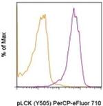
FIGURE: 1 / 1
Phospho-LCK (Tyr505) Antibody (46-9076-42) in Flow

Product Details
46-9076-42
Species Reactivity
Published species
Host/Isotype
Recommended Isotype Control
Class
Type
Clone
Immunogen
Conjugate
Excitation/Emission Max
Form
Concentration
Purification
Storage buffer
Contains
Storage conditions
Shipping conditions
RRID
Product Specific Information
Description: This SRRCHA monoclonal antibody recognizes human and mouse LCK when phosphorylated on tyrosine 505 (Y505). Y505 is a negative-regulatory site that is phosphorylated by the kinase CSK. LCK is expressed in T cells where it associates with the cytoplasmic tails of the CD4 and CD8 co-receptors on T helper cells and cytotoxic T cells, respectively. When the TCR is engaged, LCK acts to phosphorylate the intracellular chains of CD3 and zeta chains of the TCR complex, allowing the cytoplasmic tyrosine kinase, ZAP-70, to bind to them. LCK then phosphorylates and activates ZAP-70, which then phosphorylates the adaptor protein, LAT, that serves as a docking site for a number of other proteins, including SLP-76, Grb2, and phospholipase C gamma1. Mice lacking expression of LCK show defects in T cell development and function.
Applications Reported: This SRRCHA antibody has been reported for use in intracellular staining followed by flow cytometric analysis.
Applications Tested: This SRRCHA antibody has been pre-titrated and tested by intracellular staining followed by flow cytometric analysis of activated human peripheral blood cells. This can be used at 5 µL (0.06 µg) per test. A test is defined as the amount (µg) of antibody that will stain a cell sample in a final volume of 100 µL. Cell number should be determined empirically but can range from 10^5 to 10^8 cells/test.
PerCP-eFluor® 710 emits at 710 nm and is excited with the blue laser (488 nm); it can be used in place of PerCP-Cyanine5.5. We recommend using a 710/50 bandpass filter, however, the 695/40 bandpass filter is an acceptable alternative. Please make sure that your instrument is capable of detecting this fluorochrome.
Light sensitivity: This tandem dye is sensitive to photo-induced oxidation. Please protect this vial and stained samples from light.
Fixation: Samples can be stored in IC Fixation Buffer (Product # 00-8222) (100 µL of cell sample + 100 µL of IC Fixation Buffer) or 1-step Fix/Lyse Solution (Product # 00-5333) for up to 3 days in the dark at 4°C with minimal impact on brightness and FRET efficiency/compensation. Some generalizations regarding fluorophore performance after fixation can be made, but clone specific performance should be determined empirically.
Staining Protocol: All protocols work well for this monoclonal antibody. Use of Protocol A: Two-step protocol: intracellular (cytoplasmic) proteins allows for the greatest flexibility for detection of surface and intracellular (cytoplasmic) proteins. Use of Protocol B: One-step protocol: intracellular (nuclear) proteins is recommended for staining of transcription factors in conjunction with surface and phosphorylated intracellular (cytoplasmic) proteins. Protocol C: Two-step protocol: Fixation/Methanol allows for the greatest discrimination of phospho-specific signaling between unstimulated and stimulated samples, but with limitations on the ability to stain specific surface proteins (refer to "Clone Performance Following Fixation/Permeabilization" located in the Best Protocols Section under the Resources tab online). All Protocols can be found in the Flow Cytometry Protocols: "Staining Intracellular Antigens for Flow Cytometry Protocol" located in the Best Protocols Section under the Resources tab online.
Excitation: 488 nm; Emission: 710 nm; Laser: Blue Laser.
Filtration: 0.2 µm post-manufacturing filtered.
Target Information
LCK is a member of the Src-family tyrosine kinase, which are essential for signaling through the T-cell receptor (TCR) complex. Upon TCR triggering, LCK phosphorylates the ITAM motives in its zeta subunits establishing binding sites for the SH2 domains of the tyrosine kinase ZAP70, which is also phosphorylated by LCK and activated to generate subsequent signaling platforms by phosphorylation of adaptor LAT. The majority of LCK is localized to the plasma membrane, however, there is also a significant fraction associated with the Golgi apparatus which may contribute to Raf activation under conditions of weak stimulation through the TCR. LCK contains N-terminal sites for myristylation and palmitylation, a PTK domain, and SH2 and SH3 domains which are involved in mediating protein-protein interactions with phosphotyrosine-containing and proline-rich motifs, respectively. LCK is also involved in the regulation of apoptosis induced by various stimuli, but not by the death receptors. Diseases associated with LCK include Immunodeficiency 22 and CD45 deficiency. Alternatively splice variants of the LCK gene encoding different isoforms of the protein have been found.
For Research Use Only. Not for use in diagnostic procedures. Not for resale without express authorization.
How to use the Panel Builder
Watch the video to learn how to use the Invitrogen Flow Cytometry Panel Builder to build your next flow cytometry panel in 5 easy steps.
Bioinformatics
Protein Aliases: EC 2.7.10.2; kinase Lck; Leukocyte C-terminal Src kinase; LSK; Lymphocyte cell-specific protein-tyrosine kinase; OTTHUMP00000008640; OTTHUMP00000008740; OTTHUMP00000008741; p56(LSTRA) protein-tyrosine kinase; p56-LCK; Protein YT16; Proto-oncogene Lck; Proto-oncogene tyrosine-protein kinase LCK; RP4-675E8.4; T cell-specific protein-tyrosine kinase; T-lymphocyte specific protein tyrosine kinase p56lck; tyr; Tyrosine-protein kinase Lck
Gene Aliases:
Hck-3; IMD22; LCK; LSK; Lsk-t; Lskt; p56
UniProt ID: (Human) P06239, (Mouse) P06240
Entrez Gene ID: (Human) 3932, (Mouse) 16818

Performance Guarantee
If an Invitrogen™ antibody doesn't perform as described on our website or datasheet,we'll replace the product at no cost to you, or provide you with a credit for a future purchase.*
Learn more
We're here to help
Get expert recommendations for common problems or connect directly with an on staff expert for technical assistance related to applications, equipment and general product use.
Contact tech support

