Search Thermo Fisher Scientific
Invitrogen
TCR alpha/beta Monoclonal Antibody (IP26), NovaFluor™ Red 710, eBioscience™
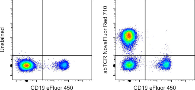
FIGURE: 1 / 2
TCR alpha/beta Antibody (H086T03R04-A) in Flow


Product Details
H086T03R04-A
Species Reactivity
Host/Isotype
Class
Type
Clone
Conjugate
Excitation/Emission Max
Form
Concentration
Purification
Storage buffer
Contains
Storage conditions
RRID
Product Specific Information
Description: The IP26 monoclonal antibody reacts with the alpha beta chain of human TCR.The alpha beta TCR is expressed by the majority of peripheral T cells.|Applications Reported: The IP26 antibody has been reported for use in flow cytometric analysis.|Applications Tested: This IP26 antibody has been pre-titrated and tested by flow cytometric analysis of human peripheral blood cells. This can be used at 5 µL (0.2 µg) per test. A test is defined as the amount (µg) of antibody that will stain a cell sample in a final volume of 100 µL. Cell number should be determined empirically but can range from 10^5 to 10^8 cells/test. Staining with the IP26 antibody can be obstructed when OKT3 (anti-human CD3) antibody is used as a co-stain.|NovaFluor dyes are not compatible with DNA intercalating viability dyes. Do not use viability dyes such as propidium iodide, 7-actinomycin D (7-AAD) and DAPI. Invitrogen LIVE/DEAD Fixable Dead Cell stains are recommended for use with NovaFluor dyes.|This NovaFluor conjugate has been updated to ship with CellBlox Plus Blocking Buffer (Cat. No. (C001T06F01)). This buffer contains formulation improvements over CellBlox. CellBlox Plus Blocking Buffer is required for optimal staining with NovaFluor conjugates and should be used in all experiments where NovaFluor conjugates are used. Whenever possible, we recommend adding CellBlox Plus Blocking Buffer to antibody cocktails/master mixes prior to combining with cells. Add 5 µL per sample (regardless of the number of NovaFluors in your panel) to use the antibody cocktail as intended. For single-color controls, use 5 µL of CellBlox Blocking Buffer per 100 µL of cell sample containing 10^3 to 10^8 cells.|NovaFluor conjugates are based on Phiton™ technology utilizing novel nucleic acid dye structures that allow for engineered fluorescent signatures with consideration for spillover and spread impacts. Learn more |Excitation: 639 nm; Emission: 710 nm; Laser: 633-640 nm (Red) Laser
Target Information
The T cell antigen receptor (TCR) consists of a ligand-specific alpha/beta heterodimer non-covalently associated with five invariant chains including the CD3 gamma/delta/eta and zeta subunits, all of which are required for efficient surface expression. T cell activation through the TCR induces cellular differentiation and/or proliferation and the production of lymphokines and cytokines. Both the CD3 and TCR zeta subunits are proposed to be responsible for the intracellular signal transduction events. Majority of T cells present in the blood, lymph and secondary lymphoid organs express TCR alpha/beta heterodimers, whereas the T cells expressing TCR gamma/delta heterodimers are localized mainly in epithelial tissues and at the sites of infection. The subunits of TCR heterodimers are covalently bonded and associate with the CD3 subunits in the endoplasmic reticulum to form functional TCR-CD3 complex. Lack of expression of any of the chains is sufficient to stop cell surface expression. The ability of T cell receptors (TCR) to discriminate foreign from self-peptides presented by major histocompatibility complex (MHC) class II molecules is essential for an effective adaptive immune response. TCR recognition of self-peptides has been linked to autoimmune disease. Mutant self-peptides have been associated with tumors. Engagement of TCRs by a family of bacterial toxins know as superantigens has been responsible for toxic shock syndrome. Autoantibodies to V beta segments of T cell receptors have been isolated from patients with rheumatoid arthritis (RA) and systemic lupus erythematosus (SLE). The autoantibodies block TH1-mediated inflammatory auto-destructive reactions and are believed to be a method by which the immune system compensates for disease (ref5). T Cell and TCR Diversity Most human T cells express the TCR alpha-beta and either CD4 or CD8 molecule (single positive, SP). A small number of T cells lack both CD4 and CD8 (double negative, DN). Increased percentages of alpha-beta DN T cells have been identified in some autoimmune and immunodeficiency disorders. Gamma-delta T cells are primarily found within the epithelium. They show less TCR diversity and recognize antigens differently than alpha-beta T cells. Subsets of gamma-delta T cells have shown antitumor and immunoregulatory activity.
For Research Use Only. Not for use in diagnostic procedures. Not for resale without express authorization.
How to use the Panel Builder
Watch the video to learn how to use the Invitrogen Flow Cytometry Panel Builder to build your next flow cytometry panel in 5 easy steps.
References (0)
Bioinformatics
Protein Aliases: FLJ22602; MGC117436; MGC22624; MGC23964; MGC71411; t-cell antigen receptor; T3/TCR complex; tcr alpha; TCR alpha/ beta; TCR beta
Gene Aliases: IMD7; TCRA; TCRB; TRA; TRB@; TRCA
Entrez Gene ID: (Human) 28755, (Human) 6957

Performance Guarantee
If an Invitrogen™ antibody doesn't perform as described on our website or datasheet,we'll replace the product at no cost to you, or provide you with a credit for a future purchase.*
Learn more
We're here to help
Get expert recommendations for common problems or connect directly with an on staff expert for technical assistance related to applications, equipment and general product use.
Contact tech support

