Search Thermo Fisher Scientific
Invitrogen
Tmem119 Monoclonal Antibody (V3RT1GOsz), PerCP-eFluor™ 710, eBioscience™
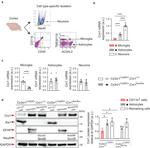
This Antibody was verified by Relative expression to ensure that the antibody binds to the antigen stated.
FIGURE: 1 / 3
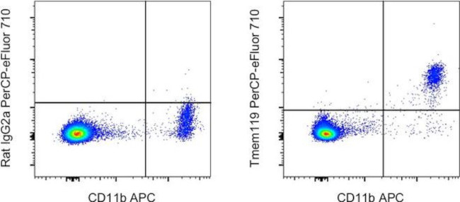
Tmem119 Antibody (46-6119-82) in Flow


Product Details
46-6119-82
Species Reactivity
Published species
Host/Isotype
Recommended Isotype Control
Class
Type
Clone
Immunogen
Conjugate
Excitation/Emission Max
Form
Concentration
Purification
Storage buffer
Contains
Storage conditions
Shipping conditions
RRID
Product Specific Information
Description: This V3RT1GOsz monoclonal antibody recognizes mouse Tmem119. This clone has been reported to be successfully used in flow cytometry.
Applications Reported: This V3RT1GOsz antibody has been reported for use in flow cytometric analysis.
Applications Tested: This V3RT1GOsz antibody has been tested by flow cytometric analysis of mouse brain cells after myelin removal. This may be used at less than or equal to 0.25 µg per test. A test is defined as the amount (µg) of antibody that will stain a cell sample in a final volume of 100 µL. Cell number should be determined empirically but can range from 10^5 to 10^8 cells/test. It is recommended that the antibody be carefully titrated for optimal performance in the assay of interest.
PerCP-eFluor 710 emits at 710 nm and is excited with the blue laser (488 nm); it can be used in place of PerCP-Cyanine5.5. We recommend using a 710/50 bandpass filter, however, the 695/40 bandpass filter is an acceptable alternative. Please make sure that your instrument is capable of detecting this fluorochrome.
Light sensitivity: This tandem dye is sensitive to photo-induced oxidation. Please protect this vial and stained samples from light.
Fixation: Samples can be stored in IC Fixation Buffer (Product # 00-8222-49) (100 µL of cell sample + 100 µL of IC Fixation Buffer) or 1-step Fix/Lyse Solution (Product # 00-5333-57) for up to 3 days in the dark at 4°C with minimal impact on brightness and FRET efficiency/compensation. Some generalizations regarding fluorophore performance after fixation can be made, but clone specific performance should be determined empirically.
Excitation: 488 nm; Emission: 710 nm; Laser: Blue Laser".
Target Information
Tmem119 encodes a single-pass type I transmembrane protein which seems to interact with a multitude of micro-RNAs as well as certain transcription factors such as SMAD1, SMAD5 and RUNX2. Tmem119 has been identified as a surface marker of microglia that can be used to reliably distinguish microglia from infiltrating macrophages. Microglial cells are phagocytes that are restricted to the brain and involved in clearing up debris and dendrite pruning of neurons. They display a high functional plasticity, balancing neuroprotective and neurotoxic functions, which is reflected by their distinct physiology in response to changes in their microenvironment. Microglia are particularly mobile in areas of the brain undergoing restructuring. Reactive microglia co-express the marker Iba1. In a pathological context, microglia have been associated with differentiation of pathogenic/inflammatory A1 type astrocytes by secretion of Cq1, IL-1a and TNF when stimulated with LPS, and thought to contribute to the pathology of several neurodegenerative diseases such as Alzheimers, Parkinsons, Chorea Huntingtons disease and Multiple Sclerosis. The specific function Tmem119 plays on microglial cells remains unclear. Outside of the neural system, Tmem119 seems to be involved in osteoblast differentiation/ossification.
For Research Use Only. Not for use in diagnostic procedures. Not for resale without express authorization.
How to use the Panel Builder
Watch the video to learn how to use the Invitrogen Flow Cytometry Panel Builder to build your next flow cytometry panel in 5 easy steps.
Bioinformatics
Protein Aliases: OBIF; Osteoblast induction factor; Transmembrane protein 119
Gene Aliases: AW208946; BC025600; obif; Tmem119
UniProt ID: (Mouse) Q8R138
Entrez Gene ID: (Mouse) 231633

Performance Guarantee
If an Invitrogen™ antibody doesn't perform as described on our website or datasheet,we'll replace the product at no cost to you, or provide you with a credit for a future purchase.*
Learn more
We're here to help
Get expert recommendations for common problems or connect directly with an on staff expert for technical assistance related to applications, equipment and general product use.
Contact tech support

