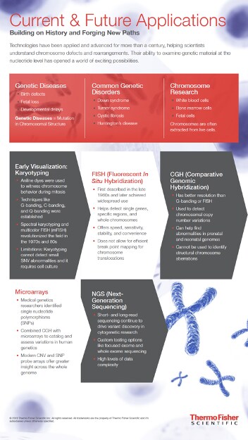
Cytogenetics has been part of the biological sciences since the mid-19th century, and this long history has meant that many techniques and methods are available to the modern cytogenetics researcher. This abundance of methods might make choosing one difficult, or make you wonder if all but the most cutting-edge approaches are functionally obsolete. In truth, each method has its own strengths and weaknesses, and choosing one depends on your research questions.

When to use chromosome banding techniques in cytogenetics research
Chromosome banding techniques were some of the first in cytogenetics and remain some of the most widely used. These early implementations of fluorescent imaging for chromosomes provide a surprising amount of information about chromosome structure. C-banding stains highly repetitive cytosine-rich regions, most often the centromeres where the chromosomal arms meet. Similarly, G-banding uses Giesma stain to mark heterochromatic regions, which are rarely transcribed and have a structural role in the overall chromosome. Q-banding uses quinacrine for similar results, but also fluoresces in proportion to the AT enrichment of the part of the chromosome to which it binds, providing more information. Each of these stains provides broad-scale information about chromosome structure, including gains, losses, translocations, and rearrangements. Thus, these techniques are useful for more than simply observing or counting chromosomes, and they remain valued diagnostic tools in medical settings. Many chromosomal abnormalities show clearly and distinctly with C, G or Q-banding, and research into these conditions often does not require more than that.
The key limitation of chromosome banding is that these methods cannot detect aberrations below 5 megabases in size, due to resolution limitations and lack of specificity. This lack of specificity also means that conditions with highly specific etiologies and research questions that need very specific data cannot be easily answered with these techniques. These methods are also limited by user error, since they are based on largely manual processing of individual microscope images, and are vulnerable to maternal cell contamination and cell culture failure.
What is fluorescence in situ hybridization (FISH)?
Fluorescence in situ hybridization (FISH) is a more tailored kind of staining. Instead of fluorescent or otherwise detectable probes binding directly to the chromosomes, probes are bound to DNA or RNA templates that then preferentially bind to their complements. This makes FISH much more specific than banding, allowing macro-scale fluorescence measurements to detect micro-scale aberrations in the chromosome. With the advent of multi-FISH, multiple fluorophores can be used in the same sample, allowing direct, in situ comparisons between differently labeled probes for multi-pronged experiments. Other variations of FISH include reverse-FISH, fiber-FISH, SKY-FISH, and more, each with its own ideal scenario.
The difficulty of FISH is that probe length is a careful balancing act. A too-short probe will have frequent off-target binding that has to be distinguished from data, but a too-long probe will face difficulties binding at all, and the line between these two scenarios is not always obvious. Like banding methods, FISH requires manual microscope image review and thus suffers from the subjectivity of its practitioners, particularly when interpreting challenging signals. FISH, in its many versions, remains common for many applications and generates striking images that are ideal for press releases and magazine covers.
What is comparative genomic hybridization?
When FISH isn’t specific enough, a related method is often the right approach: in CGH, multiple labeled DNA probes are allowed to hybridize with the sample of interest and the hybridization of each is directly compared. Typically, the probes have different labels and correspond to different versions of the same stretch of target DNA, such as different alleles or disease states. Thus, the comparison between them reveals whether the sample’s chromosomes preferentially hybridize with one or the other. With a separate, confirmed-normal control sample, it can also detect copy-number variations and translocations, including movement of a piece of the genome from one location or chromosome to another. These are all critical parts of many genetic conditions that are difficult to properly elucidate with ordinary FISH.
When to use a microarray in cytogenetics research
Microarrays are not a totally distinct method, but a different way to implement fluorescence-based genomics. These devices consist of microscope slides with thousands of tiny spots on which DNA or RNA probes are attached. The probes can be fluorescent or otherwise similar to the probes used in other techniques, including CGH and single-nucleotide polymorphism (SNP) probes. With so many probes concentrated onto a small surface, a sample can be tested for thousands of distinct molecular signatures in a single measurement, including single nucleotide polymorphisms (SNPs), copy-number variations, variant alleles, and more. Different kinds of probes have different ability to detect various signatures in a sample, and your experimental targets can make one kind of array (such as SNP) a better choice than another (such as CGH). For example, SNP arrays are much more effective at detecting long stretches of homozygosity and certain kinds of copy-number variations when compared to CGH arrays. Regardless of the kinds of probes used, the small size of each microarray well and of the overall device means that microarray output can be more easily and even automatically digitized than that of other cytogenetics methods, including in quantitative ways that are difficult to manage with other methods. Microarrays can also be purchased premade for various applications, providing considerable cost savings, and custom options exist for unusual applications that require carefully selected probes. For certain applications, microarrays are overkill, but their ease of use and ability to elucidate hundreds of diseases or disorders using a single technique has enabled them to become increasingly common in medical diagnostics, species identification, and other fields where being fast and easy is important.
In many cases, combining methods yields the best results. Although microarrays and CGH often seem like they render all other methods irrelevant, this is far from true. Microarrays often make an ideal first test that both yields extensive data and can suggest which other tests might most effectively yield more, and this approach is recommended by the American College of Medical Genetics and American College of Obstetricians and Gynecologists. Particularly vexing samples might benefit from being viewed in multiple ways, with each set of results providing context for the other. Many groups additionally use next-generation sequencing (NGS) as part of their prenatal and/or postnatal screening process, often as a second-round test to follow up on the results of other tests. As cytogenetics develops, even newer tools further expand the possibilities available to researchers, changing the mix of methods that best addresses every question and bringing ever more knowledge to light.
For more information on prenatal and postnatal genetic testing, visit our education page here
hi!,I love your writing very much! percentage we communicate extra approximately your post on AOL?
I need an expert on this space to resolve my problem.
Maybe that is you! Having a look ahead to see you.