Search Thermo Fisher Scientific
Product Specifications
Analytical sensitivity
Assay range
Sample type/volume
Hands-on time
Time-to-result
Homogenous (no wash)
Interassay CV
Intraassay CV
Instrument
Product size
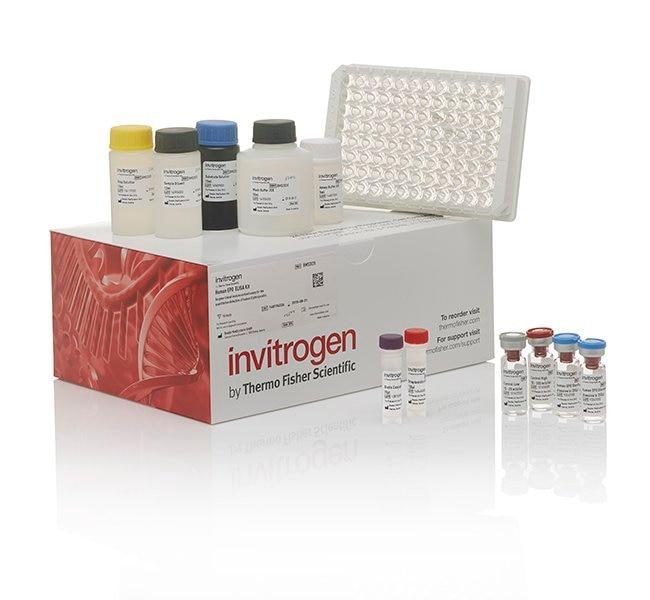
Contents
Standard
Standard Dilution Buffer
Detection Antibody
anti-rabbit-HRP
HRP Diluent
Wash Buffer
Chromogen
Stop Solution
Adhesive Plate Covers
Shipping conditions
Storage
Protein name
Target sub-specificity
Protein family
Species (tested)
Assay kit format
Detector antibody conjugate
Label or dye
About This Kit
The Human c-Myc Total (Hu c-Myc) ELISA quantitates Hu c-Myc in human cell lysates. The assay will exclusively recognize both natural and recombinant Hu c-Myc, independed of phosphorylation state.
Principle of the method
The Human c-Myc solid-phase sandwich ELISA (enzyme-linked immunosorbent assay) is designed to measure the amount of the target bound between a matched antibody pair. A target-specific antibody has been pre-coated in the wells of the supplied microplate. Samples, standards, or controls are then added into these wells and bind to the immobilized (capture) antibody. The sandwich is formed by the addition of the second (detector) antibody, a substrate solution is added that reacts with the enzyme-antibody-target complex to produce measurable signal. The intensity of this signal is directly proportional to the concentration of target present in the original specimen.
Rigorous validation
Each manufactured lot of this ELISA kit is quality tested for criteria such as sensitivity, specificity, precision, and lot-to-lot consistency. See manual for more information on validation.
The c-Myc protein is a 62 kDa transcription factor that is encoded by the c-Myc gene on human chromosome 8q24. c-Myc is commonly activated in a variety of tumor cells and plays an important role in cellular proliferation, differentiation, apoptosis and cell cycle progression. The phosphorylation of c-Myc has been investigated and previous studies have suggested a functional association between phosphorylation at Thr58/Ser62 by glycogen synthase kinase 3, cyclin dependent kinase, ERK2 and C-Jun N terminal Kinase (JNK) in cell proliferation and cell cycle regulation. Studies have shown that c-Myc is essential for vasculogenesis and angiogenesis in tumor development, which distributes blood throughout the cells. The c-myc oncogene (p62 c-myc) is involved in the control of normal cellular proliferation and differentiation, and the deregulated expression of c-Myc induces apoptosis in different cell types. Antibodies against c-myc epitopes recognize overexpressed proteins containing Myc epitope tag fused to either amino- or carboxy-termini of targeted proteins. c-Myc-, N-Myc- and L-Myc-encoded proteins function in cell proliferation, differentiation and neoplastic disease. Amplification of the c-Myc gene has been found in several types of human tumors including lung, breast and colon carcinomas, while the N-Myc gene has been found amplified in neuroblastomas. Translocation of the c-myc locus on chromosome 8 to the immunoglobulin loci on chromosome 14 (heavy chain); 2 (delta light chain); or 22 (light chain) is described in Burkitts lymphoma and other B-cell lymphoproliferative conditions. An aberrant expression of the c-myc gene occurs in tumors of different origins such as colorectal, gastric, gallbladder, hepatic, mammary, ovarian, endometrial, head and neck, pulmonary, prostatic, thyroidal, oral, ocular, nasopharyngeal, endocrine, as well as hematopoietic neoplasms.
For Research Use Only. Not for use in diagnostic procedures. Not for resale without express authorization.
Bioinformatics
Gene aliases : BHLHE39, c-Myc, MRTL, MYC, MYCC
Gene ID : (Human) 4609
Gene symbol : MYC
Protein Aliases : avian myelocytomatosis viral oncogene homolog, bHLHe39, Class E basic helix-loop-helix protein 39, Myc proto-oncogene protein, myc-related translation/localization regulatory factor, Proto-oncogene c-Myc, Transcription factor p64, v-myc myelocytomatosis viral oncogene homolog
UniProt ID (Human) P01106

Performance Guarantee
If an Invitrogen™ antibody doesn't perform as described on our website or datasheet,we'll replace the product at no cost to you, or provide you with a credit for a future purchase.*
Learn more
We're here to help
Get expert recommendations for common problems or connect directly with an on staff expert for technical assistance related to applications, equipment and general product use.
Contact tech support

