Search Thermo Fisher Scientific
Supporting cryo-EM research through reagent grants
As part of our career development and community outreach efforts at Thermo Fisher Scientific, we are excited to offer our Cryo-EM Grant Program for a second year. The program provides up to $10,000 USD of Thermo Fisher Scientific products to qualifying doctoral or post-doctoral students who are using or plan to use cryo-EM to advance their research.
2024 Cryo-EM Grant Award recipients

Dr. Katie Downes
University of Manchester
Research proposal: Visualising the early secretory pathway in situ and the hunt for mammalian COPII
Dr. Katie Downes completed her PhD in 2023 as a Presidents Doctoral Scholar under the supervision of Prof. Viki Allan at the University of Manchester, where she specialized in the cell biology of the mammalian protein secretory pathway. At least 1/3rd of our proteome uses the protein secretory pathway; defects in which are associated with a plethora of problems ranging from neurodegenerative diseases to blindness. Today, as a Postdoctoral Researcher working in the laboratory of Dr Giulia Zanetti at the Francis Crick Institute, Katie uses a suite of state-of-the-art electron and light microscopes to characterise the ultrastructure of the early secretory pathway, advancing our understanding of this vital pathway. Outside of the lab, Katie is most likely to be found surrounded by nature, be that out on a hike in the countryside or tending to her ever-growing plant collection.

Xiangsong Feng
Colombia University
Research proposal: Development of microfluidics-based time-resolved cryo-EM method for application to diverse biomolecular reaction systems
Xiangsong Feng is a postdoctoral research scientist in Dr. Joachim Frank’s lab at Columbia University in the City of New York. He obtained his Ph.D. in mechanical engineering in 2019 at Harbin Institute of Technology in China and his Ph.D. program focused on development of multi-function microfluidic chips. Fortunately, during that time, he was sponsored by a CSC fellowship from China government to visit a BioMEMS lab under Dr. Qiao Lin’s supervision in Columbia University from 2014 to 2016 and then his horizon was broadened greatly after he took a lead in developing a microspraying method in a collaborative project between Dr. Qiao Lin’s and Dr. Joachim Frank’s labs. He worked with Dr. Ziao Fu from the Dr. Frank’s lab, based on his expertise in microfluidics and the cutting-edge technique of single-particle cryo-EM from the Frank Lab, he for the first time employed the PDMS-based microspraying method to achieve a 3.0-Å structure of apoferritin. With a strong interest in the interdisciplinary field of microfluidics and single-particle cryo-EM, Xiangsong recently developed a microfluidics-based time-resolved cryo-electron microscopy (TRCEM) method to discover the dynamic process during a biological reaction before its equilibrium. As one example, this method has been successfully used for capturing on-pathway intermediates during HflX-mediated ribosome recycling, revealing a progressive splitting of the ribosome, and in this study, Xiangsong closely worked with Dr. Sayan Bhattacharjee in Dr. Frank’s lab, and he will continue to upgrade and employ the TRCEM method to study the dynamic molecular mechanism of ribosomal subunit association and the translation initiation and termination processes and many other biological reactions of interest.

Joseph Yoniles
Stanford University
Research proposal: Z-Disc Ultrastructure as the Basis of Hypertrophic Cardiomyopathy Pathogenesis
Joseph Yoniles is a second-year biophysics graduate student working between the Dunn Lab at Stanford University and the Dahlberg Lab at SLAC National Accelerator Laboratory. Early in his graduate studies, he developed a keen interest in cryogenic electron tomography (cryo-ET), captivated by the high-resolution volumetric data it provides and the complex computational methods needed to make sense of such data. Joseph is currently developing cryogenic correlative light electron microscopy (cryo-CLEM) methods to investigate the ultrastructural organization of heart muscle cells, or cardiomyocytes. He hopes that his research will enhance our understanding of the mechanisms underlying hypertrophic cardiomyopathy (HCM) and other diseases affecting heart contractility. In his free time, Joseph enjoys weightlifting, light hiking, and playing card games.
Cryo-EM Grant Program overview
Stay tuned for details on 2025 applications
The Thermo Fisher Scientific Grant Program will award three eligible doctoral or post-doctoral students with reagent awards to be used for the academic year. Applicants are asked to send in research proposals that demonstrate how they will use cryo-electron microscopy to answer their research questions. Proposals will be judged on innovative and impactful intended use of reagents and by scientific merit, significance of use, approach, and instrumentation.
The top three submissions will win:
- Gold Recipient: $10,000 USD in products
- Silver Recipient: $7,500 USD in products
- Bronze Recipient: $5,000 USD in products
Cryo-EM Grant Program submission requirements:
- Summary of your area of research (250 words max)
- Description of proposed project using cryo-EM (1,000 words max)
- References/citations to support proposed research
Thermo Fisher Scientific product awards
Winners can choose from a wide range of sample preparation products, reagents, and cryo-EM accessories.


*Cryo-EM reagents and accessories should make up 50% of the award value. Quality limits may apply for certain products.
Previous recipients
2023 Cryo-EM Grant Award recipients

Jessica Heebner
Penn State University- College of Medicine
Research proposal: Visual proteomics of neuronal growth cones using cryoET and deep learning segmentation
Jessica Heebner is a sixth year PhD candidate working in the Swulius Lab at Penn State College of Medicine studying neuron development with cryo-electron tomography. She is working towards a degree in Biomedical Science and Clinical and Translational Science. During her studies, Jess has developed a passion for image analysis and deep learning. She has developed an efficient workflow for training neural networks to segment everything from FIB-SEM to microCT to cryo-electron tomography data. Now in the final year of her program, Jess is working towards creating a whole growth cone protein atlas using a combination of montage tomography and deep learning segmentation. In her free time, Jess enjoys spending time with her pets and family and catering to the whims of a two-year-old. After graduation, she hopes to continue to develop her career in deep learning image analysis.
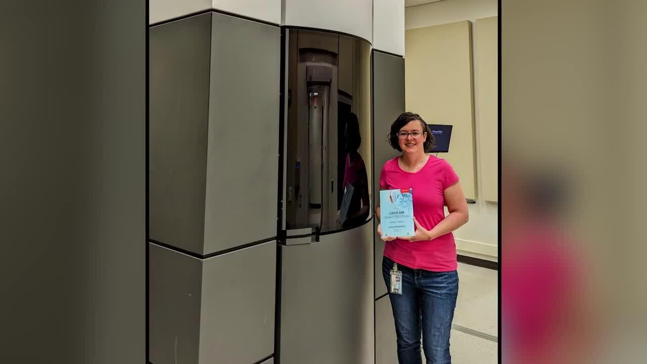

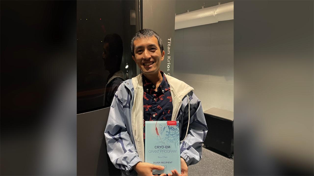
Siyu Chen
University of California, San Diego, HHMI
Research proposal: In situ structure of WT and PD mutant LRRK2 on cellular membranes
Siyu Chen is a postdoctoral fellow in the laboratory of Dr. Elizabeth Villa at UCSD, HHMI. He received his PhD in biophysics in 2022 at Northwestern University in the interdisciplinary biological sciences (IBiS) program, where he worked with Dr. Yuan He, using the cutting-edge technique of single particle cryo-EM to study the assembly and dynamics of large protein-DNA complexes and their functions in the process of DNA repair and transcription initiation. Siyu’s PhD training was sponsored by the Rappaport award from the department and the Molecular Biophysics training grant from NIH.
With a solid training in the field of structural biology on single-particle cryo-EM, Siyu aims to extend his research interests into in situ structural biology during his postdoctoral tenure, obtaining high-resolution maps of molecular assemblies when they function in their native environment. In Dr. Elizabeth Villa’s biophysics and cellular biology laboratory, he will utilize the state-of-the-art techniques of cryo-electron tomography with focused ion-beam milling (cryo-FIB-ET) to study the molecular mechanism of LRRK2 function at lysosomes and Golgi networks, as well as how mutant LRRK2 is related to Parkinson’s disease. With his background and enthusiasm, as well as the leading-edge technologies being developed in close collaboration with Thermo Fisher Scientific, Siyu will devote himself to help advance the understanding of molecular mechanisms of PD and contribute to our fight against this disease.

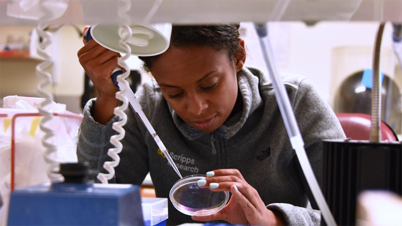
Lisa Eshun-Wilson
Scripps Research Institute
Research proposal: Uncovering the molecular mechanisms of mitochondrial proteostasis
Dr. Eshun-Wilson is currently a National Science Foundation Postdoctoral Fellow (NSF-PRFB) in the laboratory of Dr. Gabriel Lander at the Scripps Research Institute in La Jolla. She completed her PhD in the laboratory of Dr. Eva Nogales at UC Berkeley as a recipient of the National Science Foundation Graduate Research Fellowship Program (NSF-GRFP) and Ford Foundation Fellowship. She also received the 2020 Cris Alvaro PhD Commencement Prize for excellence in research and community service. Currently, she uses the most powerful microscopes on the planet, transmission electron microscopes, to visualize the ways molecular machines can be harnessed to fight disease, with an emphasis on mitochondrial quality control proteases—or, in other words, vital stress sensors of the cell. She aims to characterize how these biological motors communicate within the body, providing avenues for therapeutic intervention in cancer, heart disease, and neurodegeneration.
For Research Use Only. Not for use in diagnostic procedures.