Search Thermo Fisher Scientific
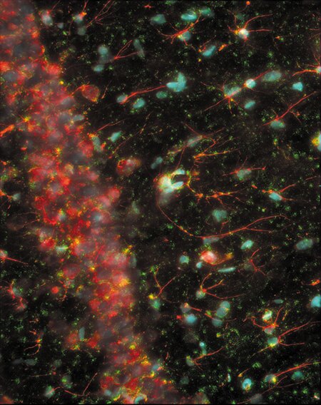
Immunohistochemistry using GFAP Monoclonal Antibody, Mouse, Alexa Fluor® 594 Conjugate
Immunohistochemistry using NMDA Receptor Subunit 2A Polyclonal Antibody, Rabbit: Rat brain cryosections labeled with anti–NMDA receptor, subunit 2A (rat) antibody, rabbit IgG fraction (A6473) and detected using Alexa Fluor 488 goat anti–rabbit IgG antibody (A11008). The tissue was also labeled with Alexa Fluor 594 anti–glial fibrillary acidic protein antibody (A21295) and counterstained with TOTO-3 iodide (T3604), which was pseudocolored light blue in this image.

Related Products
Related Images
A prometaphase muntjac skin fibroblast stained with Alexa Fluor® 350 phalloidin, an anti–a-tubulin antibody and an anti–cdc6 peptide antibody. Go ›

Bovine pulmonary artery endothelial cells (BPAEC). MitoTracker® Red CMXRos, SYTOX® Green nucleic acid stain, biotin-XX goat anti–mouse IgG antibody and Cascade Blue® NeutrAvidin biotin-binding protein. Go ›

1% Agarose gel containing 16S and 23S ribosomal RNA (rRNA). SYBR® Green II RNA gel stain. Go ›

Mouse Anti-Alpha Tubulin Monoclonal Antibody (Cat. No. A11126) Go ›

CD335 (NKp46) Antibody (63335182) in RE Go ›

CD223 (LAG-3) Antibody (56223942) in TM Go ›

Increase in accuracy and precision using the Qubit® Go ›

Performance of the Qubit® dsDNA HS Assay Go ›

Developing Drosophila embryo Go ›

Cytoskeleton of a mixed population of granule neurons and glial cells Go ›

Muntjac fibroblast labeled with probes for actin and the nucleus. Go ›

Multicolor fluorescence analysis of muntjac fibroblasts. Go ›
