Search Thermo Fisher Scientific
B Cell Activation Functional Assay
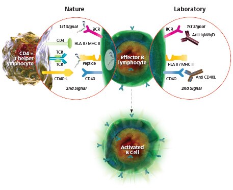
B cell activation, like T cell activation, also requires two signals. The first signal is provided by the B Cell Receptor (BCR), a surface-expressed antibody binding to its cognate antigen. This signal may also be mimicked using anti-IgM or IgD antibodies. The second signal is achieved through engagement of co-stimulatory molecules such as CD40 and cytokine signaling. Alternatively, components of bacterial cell walls, such as Lipopolysaccharide (LPS), and antigens with highly repetitious molecules may signal B cell activation directly.


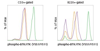
Mouse splenocytes were left untreated or were activated with anti-mouse IgM, u chain specific, antibody or with sodium pervanadate. Cells were then surface stained with anti-CD3 and with anti-B220 and intracellularly stained with anti-phospho-BTK/ITK (M4G3LN) PCP-eFluor® 710. Histogram shows levels of phospho-BTK/ITK on CD3+-gated T cells (left) and B220+-gated B cells (right). Orange = unstimulated, purple = +a-IgM, green = +Na3VO4.
For Research Use Only. Not for use in diagnostic procedures.