Search Thermo Fisher Scientific
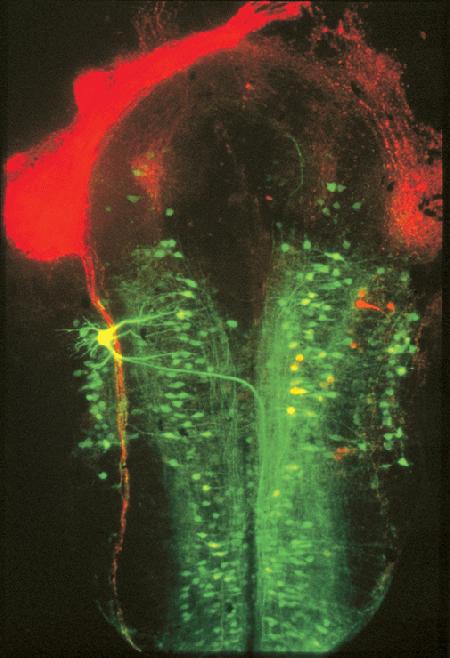
Brain of a Xenopus laevis embryo.
A whole mount of the embryonic brain of Xenopus laevis that has been double-labeled with our lysine-fixable 10,000 MW fluorescein dextran (fluoro-emerald, Cat. No. D1820) and lysine-fixable 10,000 MW tetramethylrhodamine dextran (fluoro-ruby, Cat. No. D1817). The tetramethylrhodamine dextran was used to label the neurons projecting from the retina, whereas the fluorescein dextran was applied to the transected spinal cord, thus allowing the detailed evaluation of the topological relationship of these two populations of neurons. Image contributed by Martina Manns and Bernd Fritzsch, Creighton University.

