Search Thermo Fisher Scientific
Two-photon excitation imaging of calcium influx in a CA1 pyramidal cell spine
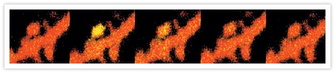
Two-photon excitation imaging of Ca2+ influx in a CA1 pyramidal cell spine. The images are overlays of anatomical images generated by Alexa Fluor® 594 hydrazide (Cat. no. A10438, A10442) and Ca2+ signals generated by fluo-5F (Cat. no. F14221). The imaging system uses two-photon excitation at 810 nm and two-channel emission detection (fluo-5F in the green channel, Alexa Fluor® 594 hydrazide in the red channel). The observed Ca2+ influx is through NMDA receptors that are activated by glutamate released from the presynaptic terminal following electrode stimulation of a collateral CA3 pyramidal cell axon. The brief (0.2 ms) depolarizing stimulus was applied after the first frame in the image sequence. The frame rate is four frames/second, and each frame represents an area of 5 µm × 5 µm. The image was contributed by Thomas Oertner and Karel Svoboda, Cold Spring Harbor Laboratory.

Related Products
Related Images
REF-52 fibroblasts. Cyclic AMP Fluorosensor (FlCRhR) and fura-2 AM Go ›

Pseudocolored images of changes in intracellular free Ca2+ in AtT-20/D16v-F2 cells, monitored at 9 sec intervals with fluo-4, AM (F14201, F14202, F14217, F23917). Go ›

Cryostat section of mouse kidney. FluoCells® prepared slide #3, Alexa Fluor® 488 wheat germ agglutinin, Alexa Fluor® 568 phalloidin and DAPI. Go ›

CA1 pyramidal neuron in a hippocampal slice filled with SBFI delivered from a patch pipette. Go ›
