Search Thermo Fisher Scientific
Invitrogen
CD134 (OX40) Monoclonal Antibody (OX-86), PerCP-eFluor™ 710, eBioscience™
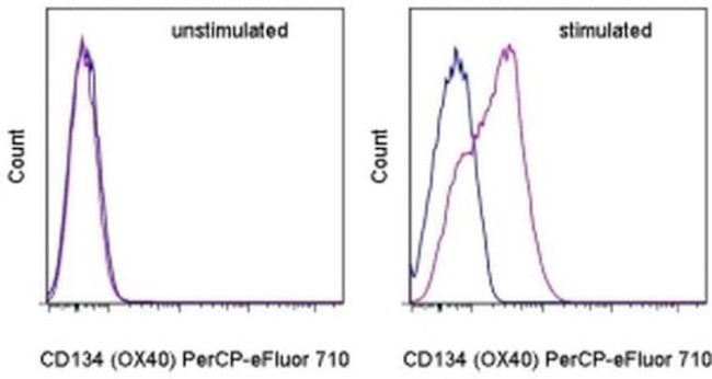
FIGURE: 1 / 3
CD134 (OX40) Antibody (46-1341-82) in Flow


Product Details
46-1341-82
Species Reactivity
Published species
Host/Isotype
Recommended Isotype Control
Class
Type
Clone
Conjugate
Excitation/Emission Max
Form
Concentration
Purification
Storage buffer
Contains
Storage conditions
Shipping conditions
RRID
Product Specific Information
Description: The OX-86 monoclonal antibody reacts with mouse CD134, also known as OX40. A member of the TNF receptor superfamily, CD134 is a 50 kDa type I membrane glycoprotein expressed by activated mouse T lymphocytes. Rat CD134 was initially identified as an activation marker only on activated CD4+ T cells. In contrast, mouse CD134 is expressed by both activated CD4+ and CD8+ T cells. The interaction of CD134 with CD252 (OX40 ligand) has been implicated in T cell-dependent humoral responses, regulation of primary T cell expansion, survival of T cells, size of the memory T cell pool, and regulation of tolerance in the CD4+ T cell compartment.
Applications Reported: This OX-86 antibody has been reported for use in flow cytometric analysis.
Applications Tested: This OX-86 antibody has been tested by flow cytometric analysis of stimulated mouse splenocytes. This can be used at less than or equal to 0.25 µg per test. A test is defined as the amount (µg) of antibody that will stain a cell sample in a final volume of 100 µL. Cell number should be determined empirically but can range from 10^5 to 10^8 cells/test. It is recommended that the antibody be carefully titrated for optimal performance in the assay of interest.
PerCP-eFluor® 710 emits at 710 nm and is excited with the blue laser (488 nm); it can be used in place of PerCP-Cyanine5.5. We recommend using a 710/50 bandpass filter, however, the 695/40 bandpass filter is an acceptable alternative. Please make sure that your instrument is capable of detecting this fluorochrome.
Fixation: Samples can be stored in IC Fixation Buffer (Product # 00-822-49) (100 µL of cell sample + 100 µL of IC Fixation Buffer) or 1-step Fix/Lyse Solution (Product # 00-5333-54) for up to 3 days in the dark at 4°C with minimal impact on brightness and FRET efficiency/compensation. Some generalizations regarding fluorophore performance after fixation can be made, but clone specific performance should be determined empirically.
Excitation: 488 nm; Emission: 710 nm; Laser: Blue Laser.
Filtration: 0.2 µm post-manufacturing filtered.
Target Information
OX40 is a protein receptor found on the surface of T cells, a type of white blood cell that plays a crucial role in the immune system. When activated, OX40 promotes the proliferation and survival of T cells, leading to a stronger immune response against cancer cells and infectious agents. OX40 agonists, which are drugs that activate OX40, have shown promising results in preclinical and clinical studies as a potential immunotherapy for cancer. They can enhance the efficacy of other cancer treatments such as chemotherapy and checkpoint inhibitors, and may also have applications in autoimmune diseases and allergies.
For Research Use Only. Not for use in diagnostic procedures. Not for resale without express authorization.
How to use the Panel Builder
Watch the video to learn how to use the Invitrogen Flow Cytometry Panel Builder to build your next flow cytometry panel in 5 easy steps.
Bioinformatics
Protein Aliases: CD134; OX40 antigen; OX40L receptor; tax-transcriptionally activated glycoprotein 1; Tumor necrosis factor receptor superfamily member 4
Gene Aliases: ACT35; CD134; Ly-70; Ox40; Tnfrsf4; Txgp1; TXGP1L
UniProt ID: (Mouse) P47741
Entrez Gene ID: (Mouse) 22163

Performance Guarantee
If an Invitrogen™ antibody doesn't perform as described on our website or datasheet,we'll replace the product at no cost to you, or provide you with a credit for a future purchase.*
Learn more
We're here to help
Get expert recommendations for common problems or connect directly with an on staff expert for technical assistance related to applications, equipment and general product use.
Contact tech support

