Search Thermo Fisher Scientific
Invitrogen
CD63 Monoclonal Antibody (H5C6), eFluor™ 450, eBioscience™
FIGURE: 1 / 9
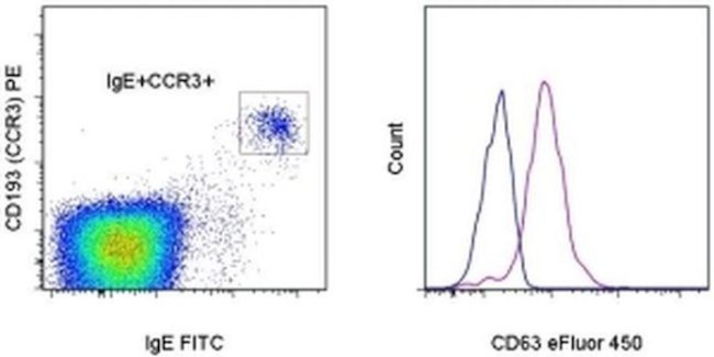
CD63 Antibody (48-0639-42) in Flow




Product Details
48-0639-42
Species Reactivity
Published species
Host/Isotype
Recommended Isotype Control
Class
Type
Clone
Conjugate
Excitation/Emission Max
Form
Concentration
Purification
Storage buffer
Contains
Storage conditions
Shipping conditions
RRID
Product Specific Information
Description: This H5C6 monoclonal antibody reacts with human CD63, a type III member of the tetraspanin family of transmembrane proteins. CD63 is expressed intracellularly on lysosomes, endosomes, and granules of resting platelets and basophils. However, cell surface expression of CD63 can be detected on activated basophils and platelets, monocytes, macrophages, and granulocytes. This receptor is also expressed on endothelial cells, fibroblasts, and smooth muscle cells. Studies have demonstrated that CD63 associates with integrins (VLA-3 and VLA-6) and TIMP-1 to mediate the allergic response.
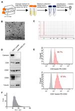
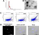
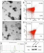
Applications Reported: This H5C6 antibody has been reported for use in flow cytometric analysis.
Applications Tested: This H5C6 antibody has been pre-titrated and tested by flow cytometric analysis of normal human peripheral blood cells. This can be used at 5 µL (1 µg) per test. A test is defined as the amount (µg) of antibody that will stain a cell sample in a final volume of 100 µL. Cell number should be determined empirically but can range from 10^5 to 10^8 cells/test.
eFluor® 450 is an alternative to Pacific Blue®. eFluor® 450 emits at 445 nm and is excited with the Violet laser (405 nm). Please make sure that your instrument is capable of detecting this fluorochome.
Excitation: 405 nm; Emission: 445 nm; Laser: Violet Laser.
Filtration: 0.2 µm post-manufacturing filtered.
Target Information
CD63 (LAMP-3, lysosome-associated membrane protein-3), a glycoprotein of tetraspanin family, is present in late endosomes, lysosomes and secretory vesicles of various cell types. CD63 is also present in the plasma membrane, usually following cell activation. Hence, CD63 has become a widely used basophil activation marker. In mast cells, however, CD63 exposition does not need their activation. CD63 interacts with integrins and affects phagocytosis and cell migration, it is also involved in H/K-ATPase trafficking regulation of ROMK1 channels. CD63 also serves as a T-cell costimulation molecule. Expression of CD63 can be used for predicting the prognosis in earlier stages of carcinomas. CD63 is expressed on activated platelets, and is a lysosomal membrane glycoprotein that is translocated to plasma membrane after platelet activation. CD63 is also present in monocytes and macrophages and is weakly expressed on granulocytes, B, and T cells. CD63 is identical to the melanoma-associated antigen which is ME491 and to the platelet antigen PTLGP40. Diseases associated with CD63 dysfunction include melanoma and Hermansky-Pudlak Syndrome.
For Research Use Only. Not for use in diagnostic procedures. Not for resale without express authorization.
How to use the Panel Builder
Watch the video to learn how to use the Invitrogen Flow Cytometry Panel Builder to build your next flow cytometry panel in 5 easy steps.
Bioinformatics
Protein Aliases: CD 63; CD63; CD63 antigen; CD63 antigen (melanoma 1 antigen); Granulophysin; LAMP-3; Limp1; Lysosomal-associated membrane protein 3; Lysosome integral membrane protein 1; lysosome-associated membrane glycoprotein 3; Melanoma-associated antigen ME491; melanoma-associated antigen MLA1; Ocular melanoma-associated antigen; OMA81H; Tetraspanin-30; Tspan-30
Gene Aliases: CD63; LAMP-3; ME491; MLA1; OMA81H; TSPAN30
UniProt ID: (Human) P08962
Entrez Gene ID: (Human) 967

Performance Guarantee
If an Invitrogen™ antibody doesn't perform as described on our website or datasheet,we'll replace the product at no cost to you, or provide you with a credit for a future purchase.*
Learn more
We're here to help
Get expert recommendations for common problems or connect directly with an on staff expert for technical assistance related to applications, equipment and general product use.
Contact tech support
