Search Thermo Fisher Scientific
- Order Status
- Quick Order
-
Don't have an account ? Create Account
Search Thermo Fisher Scientific
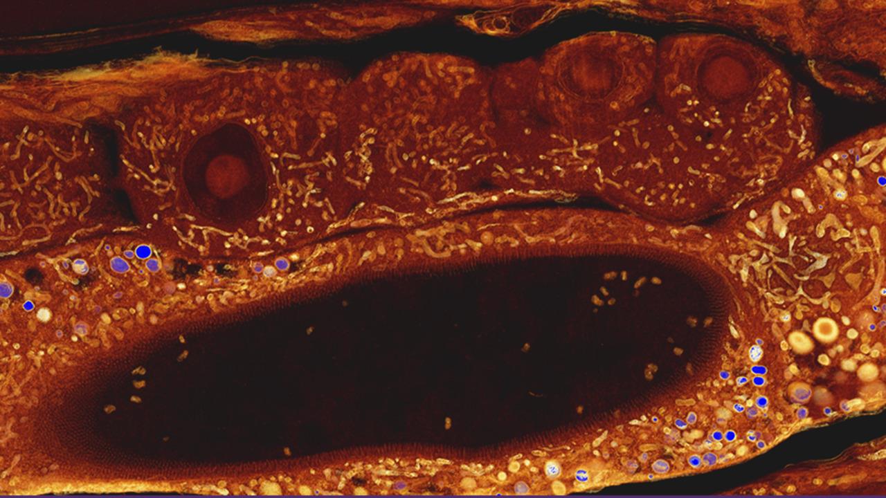
Array tomography can provide rapid localization of cells and their interaction partners in tissues. With the largest imaging field, array tomography is ideal for tissue histology. Ultrastructure analysis of whole animals or whole organs is not needed, so array tomography provides easy navigation for samples that can be stored and re-imaged.
Turn any Thermo Scientific SEM into a volume EM microscope with the addition of automated Array tomography.
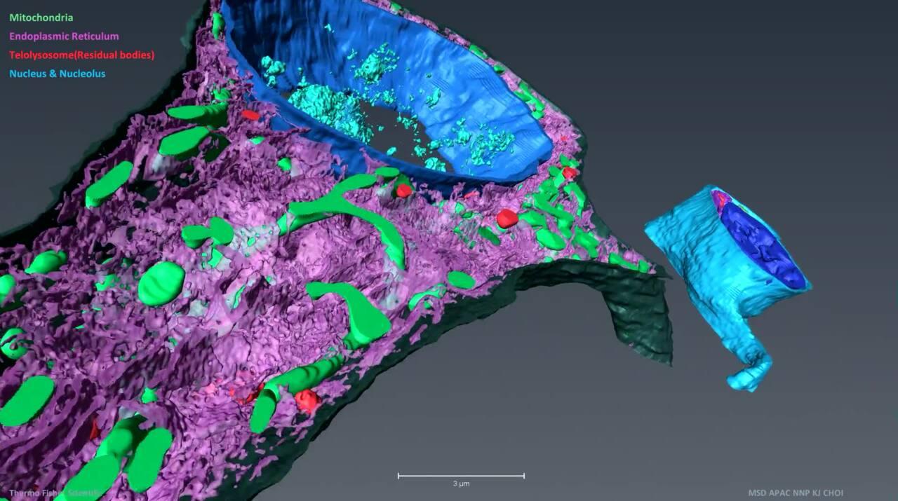
 Array tomography analysis of a mouse brain
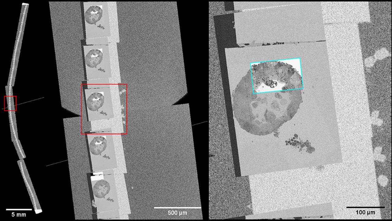
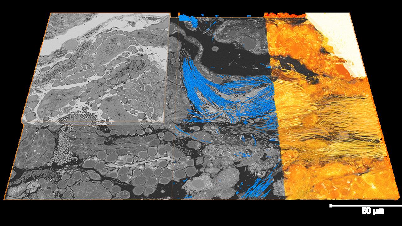
Thermo Scientific Maps Correlative Electron Microscopy Software guides the acquisition of 3D volumes for array tomography. When a user employs an SEM-based method for 3D reconstruction, the total acquisition time can quickly become impractical if the selected volume is unnecessarily large. In that case, the ability to precisely identify an area of interest becomes particularly important. Given the capacity to create large overviews at high resolution and the correlation with fluorescent images, Maps provides the 2D map to target 3D acquisitions (e.g., using Thermo Scientific Auto Slice & View Software), thus delivering results more quickly.
Additionally, the modular architecture of Maps software allows integration of more complex, SEM-based, 3D acquisition schemes. Maps software allows users to define multiple imaging areas on the block face and guides the user through the setup of slicing and imaging parameters. With the optional array tomography plugin for SEM/DualBeam systems, Maps software assists the user in imaging serial sections for 3D reconstruction.


“In Maps with array tomography, less than 10µm section-to-section variability of imaging region placement allows imaging with much less overhead. This variability can be hundreds of microns in other solutions. Imaging with less overhead speeds up time to data by a large factor especially when imaging small target structures.”
Narayanan 'Bobby' Kasthuri, Neuroscientist, University of Chicago

“Electron microscopy analysis is frequently complex. Our array tomography protocol with Maps provides a more straightforward electron microscopy data analysis. This facilitates processing and acquisition of the EM data from the large sample surface and intermediate volume. Given the overall ease of the approach, we believe that it can be used to address not only scientific questions, but also as a diagnostic tool.”
Irnia Kolotuev, EM Facility Coordinator, University of Lausanne
For Research Use Only. Not for use in diagnostic procedures.