Search Thermo Fisher Scientific
- Order Status
- Quick Order
-
Don't have an account ? Create Account
Search Thermo Fisher Scientific
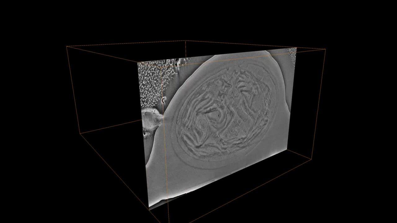
A plasma focused ion beam milling scanning electron microscopy (plasma-FIB SEM) combines improved sputtering efficiency with nanometer imaging resolution at either room temperature or under cryogenic conditions. This can provide fast-end pointing during 3D imaging. Multiple ion species (Xe, O, Ar, and N) are versatile options for site-specific, large-volume material removal. For example, O+ PFIB provides superior data acquisition efficiency and image quality for samples embedded in either epoxy- or acrylic-based resins. Unlike Ga-based FIBs, curtain free surfaces are easily generated for a wide range of materials such as LR-White, HM20, and EPON resins. Superior resin- and sample-processing compatibility allows for targeted FIB-SEM tomography with direct, in-resin correlative imaging of the region of interest (ROI). ROI targeting can be further enhanced by correlative light and electron microscopy (CLEM). Data management of slices is automated.
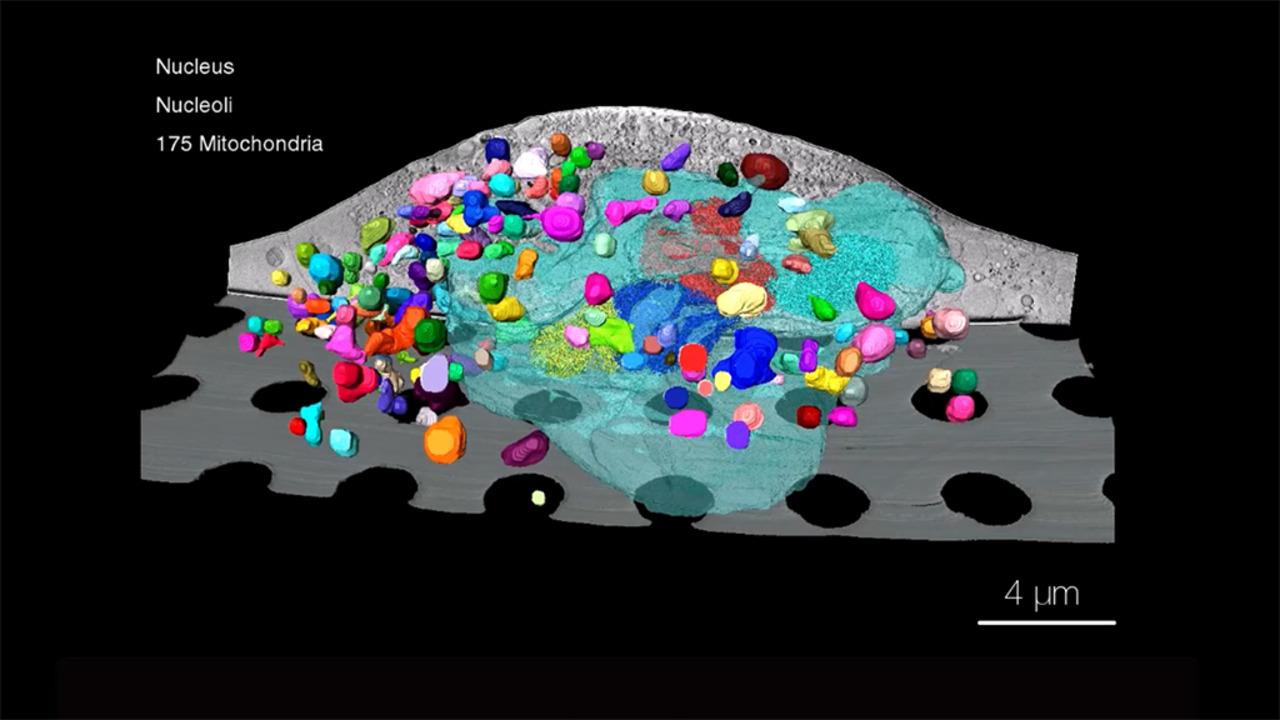
 3D volume of mouse neuron containing synaptic vesicles and mitochondria (colored) HPF in EPON.
The Thermo Scientific Hydra Bio Plasma-FIB is a versatile tool for high-throughput cellular FIB-SEM tomography, compatible with all commonly used sample-embedding media and preparation protocols. Four ion species (Xe, O, Ar, and N) can be used independently for site-specific, large-volume material removal for top-down and cross-section analysis in 2D and 3D. By selecting the ion beam that matches the requirements of each individual sample, excellent surface texture can be achieved for complex samples, including sample-substrate interfaces and dental (mineralized tissue) materials.
A plasma FIB enables efficient, large-volume serial sectioning thanks to a broader, collimated beam delivering currents up to 2.5 μA higher than a Ga-FIB. Improved sputtering efficiency enhances performance, generating smoother cut faces and reducing curtaining artifacts, further improving throughput, and providing fast access to regions of interest. The combination of higher currents, higher sputter rates, and reduced damage makes it possible to access volumes hundreds of micrometers in size while still observing nanoscale features.

The optional Spin Mill Bio Method on the Hydra Bio Plasma-FIB provides large-area planar milling up to 1 mm in diameter and the ability to image large areas in a horizontal plane for 3D characterization. The Spin Mill process is fully automated and easy to set up using Thermo Scientific Auto Slice & View Software. Multiple areas for image acquisition within one Spin Mill Bio experiment can be selected. Each area of interest can be imaged at different imaging settings based on the specificity of the experiment.
With Spin Mill, samples are milled at a near-glancing angle with the PFIB; typical sample preparation for slice-and-view analysis (e.g., protective capping, trenching, or the use of fiducial marks) is not required. With such a large, milled area, numerous regions can be selected and imaged. Sparse features can easily be identified, and statistically relevant 3D data can be collected from multiple areas.

The Thermo Scientific iFLM Correlative System is an integrated wide-field light microscope for cryo-correlative imaging inside the Hydra Bio Plasma-FIB allowing you to combine fluorescence imaging and ion milling within a single microscope for correlative light and electron microscopy (CLEM) at room temperature or under cryo conditions.


“We are increasingly being challenged to understand how to improve crop resilience to environmental stress and disease. The remarkable capabilities of our new Hydra vEM will allow us the unprecedented ability to reconstruct entire plant cells and tissue with exquisite detail. This technology will allow us to “freeze” organisms in time and space and build intricate 3D models that will help us solve our critical food security challenges.”
Kirk J. Czymmek, Principal Investigator, Director Advanced Bioimaging Laboratory, Donald Danforth Plant Science Center
For Research Use Only. Not for use in diagnostic procedures.