Support for Countess II Automated Cell Counters: online and downloadable materials

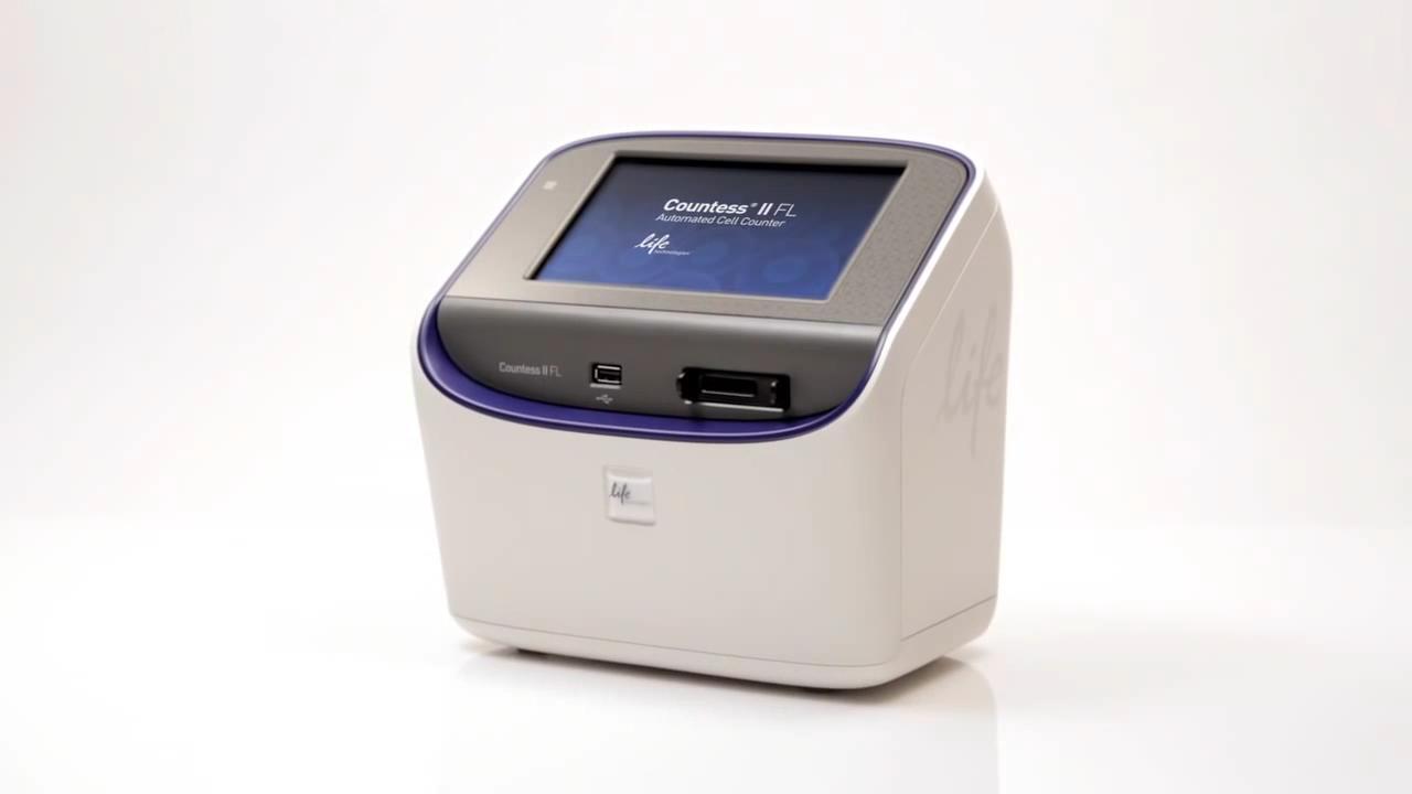
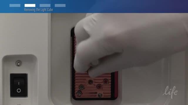
The manuals, brochures, application notes, downloads, demos, videos, and other resources in this section will help you get the most out of your Countess II Automated Cell Counter.


Documentation and literature


Customer stories

“Fast count and nice instrument, easy to use!! I love the small counter!”

Dr. Xin Xu, Baylor College of Medicine

“It’s very easy to use and it also allows me to quickly update my lab notebook so that each experiment uses the same number of cells. Quick and efficient.”

Lakeisha Tillery, Meharry Medical College

“It outperformed all other competitors' models that we tried.”

Michael DaCosta, The Jackson Laboratory

“It is very effective at counting cells. It works very well.”

Mairead Bretney, Sanford Burnham Medical Research Institute

“The Countess II makes it very easy to count cells accurately and quickly—the fact that it tells you the dead cell and live cell count is icing on the cake.”

Faryal Mir, MIT

“We compared to a hemocytometer and the Countess II always obtained very close results. The fluorescent measurements are a great tool for us to quantify the % transfection and transduction efficiency in our experiments. The light cubes used to detect fluorescence are very easy to switch out when more than two fluorescence sources are needing to be detected. The automatic focusing is very effective and accurate with the option to manually focus.”

David Waynar, Santa Cruz Biotechnology

“This equipment is very useful for the purposes it is used in our lab. We love it!”

Kristin Harwood, BioMarker Strategies, LLC

“Great for a quick check of transfection/transduction efficiency. Easy to use and seems pretty sensitive.”

Alex Peister, Morehouse College

“Great device. Broad range of cell densities that it can handle. Good at distinguishing clumps and counting cells within clumps.”

Joanne Rzepka, Veterinary Medical Research & Development

“Low maintenance, fast easy accurate cell counts. Easy for students to use and comprehend.”

Robert Dean, D’Youville College

“A very accurate cell counter and you can use it to count fluorescent cells as well. Excellent product. I’m very happy with it. I highly recommend it.”

Massimo Ammirante, University of California, San Diego


Videos


Software updates

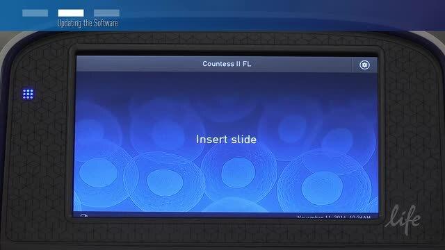
To download the latest software for Countess II Automated Cell Counters, see our Software Download page. Then view the software update how-to video in the Videos section of this page.

Educational resources

Educational resources for Countess Automated Cell Counters include an abundance of materials on cell imaging, cell counting, and cell culture. For training courses taught at your site and online, see Services.

Cell Analysis Learning Center

The learning center contains resources for cell analysis techniques and basic principles. Subtopics relevant to Countess II products include Cell counting information and the Molecular Probes School of Fluorescence.

Gibco Cell Culture Basics

Our free introduction to cell culture, covering topics such as laboratory setup, safety, and aseptic technique. You’ll also find basic cell culture techniques and methods for passaging, freezing, and thawing cultured cells.

Verified cell lines

Cell lines verified on the Countess II Automated Cell Counters


Citations

Shi B et al. (2018). Inhibition of HIV early replication by the p53 and its downstream gene p21. Virology journal 15(1) 53.


Sykora P et al. (2018). Next generation high throughput DNA damage detection platform for genotoxic compound screening. Scientific reports 8(1) 2771.


Elango J et al. (2018). Cross-talk between primary osteocytes and bone marrow macrophages for osteoclastogenesis upon collagen treatment. Scientific reports 8(1) 5318.


Poh A R et al. (2017). Inhibition of hematopoietic cell kinase activity suppresses myeloid cell-mediated colon cancer progression. Cancer cell 31(4) 563–575.


Feng Y et al. (2016) Ratiometric electrochemiluminescence detection of circulating tumor cells and cell-surface glycans. J Electroanal Chem 25 July 2016.


Pirhaji L et al. (2016) Revealing disease-associated pathways by network integration of untargeted metabolomics. Nat Methods 13(9):770–776.doi:10.1038/nmeth.3940.


Delgado RN et al. (2016) Maintenance of neural stem cell regional identity in culture. Neurogenesis 3(1).


Murtaza M et al. (2016) Rotenone susceptibility phenotype in olfactory derived patient cells as a model of idiopathic Parkinson’s Disease. PLoS One 11(4):e0154544. doi:10.1371/journal.pone.0154544.


Vuković LD et al. (2016) Nuclear size is sensitive to NTF2 protein levels in a manner dependent on Ran binding. J Cell Sci 129(6):1115–1127.


Jang DH et al. (2016) Impairment of mitochondrial respiration following ex vivo cyanide exposure in peripheral blood mononuclear cells. Clin Toxicol (Phila) 54(4):303–307.


Jiang X et al. (2016) Protective effect of C4a against hyperoxic lung injury via a macrophage‑dependent but not a neutrophil/lymphocyte‑dependent signaling pathway. Mol Med Rep 13(2):1250–1256.


Wu R et al. (2015) Fabrication and evaluation of multilayer nanofiber-hydrogel meshes with a controlled release property. Fibers 3(3):296–308.


Labedz-Maslowska A et al. (2015) Monocyte chemoattractant protein-induced protein 1 (MCPIP1) enhances angiogenic and cardiomyogenic potential of murine bone marrow-derived mesenchymal stem cells. PLoS One 10(7):e0133746.


Huynh LM et al. (2015) Two histone variants TH2A and TH2B enhance human induced pluripotent stem cell generation. Stem Cells Dev 25(3):251–258.

Takahashi T (2018). Applicability of automated cell counter with a chlorophyll detector in routine management of microalgae. Scientific reports 8(1) 4967.


Jimenez A G et al. (2018) Effects of short‐term clomipramine on anxiety‐like behavior cellular metabolism and oxidative stress in primary fibroblast cells of male and female rats. Physiological reports 6(9) e13615.


Dyavar S R et al. (2018). Normalization of cell associated antiretroviral drug concentrations with a novel RPP30 droplet digital PCR assay. Scientific reports 8(1) 3626.


Li H et al. (2018). Antitumor activity of EGFR-specific CAR T cells against non-small-cell lung cancer cells in vitro and in mice. Cell death & disease 9(2) 177.


Dong L et al. (2018). Verteporfin inhibits YAP-induced bladder cancer cell growth and invasion via Hippo signaling pathway. International journal of medical sciences 15(6) 645.


DeRosa B A et al. (2018). Convergent pathways in idiopathic autism revealed by time-course transcriptomic analysis of patient-derived neurons. Scientific Reports 8(1) 8423.


Akatsuka A et al. (2016) A novel thiophene‐3‐carboxamide analog of annonaceous acetogenin exhibits antitumor activity via inhibition of mitochondrial complex I. Pharmacol Res Perspect 4(4)


Meng XQ et al. (2016) Solanine-induced reactive oxygen species inhibit the growth of human hepatocellular carcinoma HepG2 cells. Oncol Lett 11(3):2145–2151


Huang CH et al. (2016) Paper-based microreactor array for rapid screening of cell signaling cascades. Lab Chip 16(15):2911–2920


Swioklo S et al. (2016) Alginate-encapsulation for the improved hypothermic preservation of human adipose-derived stem cells. Stem Cells Transl Med 5(3):339–349.


Huang D et al. (2016) Rapid separation of human breast cancer cells from blood using a simple spiral channel device. Anal Methods 8(30):5940–5948.


Choudhury B et al. (2016) Anticancer activity of Garcinia morella on T-cell murine lymphoma via apoptotic induction. Front Pharmacol 7:3


Yang H et al. (2016) Effect of a feeder layer composed of mouse embryonic and human foreskin fibroblasts on the proliferation of human embryonic stem cells. Exp Ther Med 11(6):2321–2328.


MacLean JA II et al. (2016) WNT7A regulation by miR-15b in ovarian cancer. PLoS One 11(5):e0156109. doi:10.1371/journal.pone.0156109.


Wang J et al. (2016) Investigation of the anti-asthmatic activity of Oridonin on a mouse model of asthma. Mol Med Rep 14(3):2000–2006


Deleon H (2016) Application of polystyrene disk substrates in cellular cultivation methods: generalized specimen preparation protocol for scanning electron microscopy. J Microsc May 30. doi:10.1111/jmi.12429.


Hasegawa M et al. (2016) Surface coating of a metal mesh device sensor with gold to improve the separation and sensing of mammalian cells. IEEE Sens J 16(13):5129–5135.


Ok SH et al. (2016) Lipid emulsion attenuates apoptosis induced by a toxic dose of bupivacaine in H9c2 rat cardiomyoblast cells. Human Exp Toxicol 35(9):929–937. doi:10.1177/0960327115608930.


Li K et al. (2015) Clinically applicable magnetic-labeling of natural killer cells for MRI of transcatheter delivery to liver tumors: preclinical validation for clinical translation. Nanomedicine (Lond) 10(11):1761–1774.


Roma A et al. (2015) Selective induction of apoptosis by Azadirachta indica leaf extract by targeting oxidative vulnerabilities in human cancer cells. J Pharm Pharm Sci 18(4):729–746.


Malvicini M et al. (2015) Tumor microenvironment remodeling by 4-methylumbelliferone boosts the antitumor effect of combined immunotherapy in murine colorectal carcinoma. Mol Ther 23(9):1444–1455.


Han X et al. (2015) CRISPR-Cas9 delivery to hard-to-transfect cells via membrane deformation. Sci Adv 1(7):e1500454


Register your instrument

Register your instrument to receive notifications of software and firmware updates.


For Research Use Only. Not for use in diagnostic procedures.