Search Thermo Fisher Scientific
Electron microscopy imaging and analysis resources for batteries
Battery imaging and analysis webinars

Webinar: Characterization of batteries and energy materials: Avizo Software
In this introductory webinar, you will learn how to use Thermo Scientific Avizo Software workflows to repeatably visualize and analyze battery samples from 2D and 3D acquisition techniques. Identify and measure complex features in electrode materials and entire cells with high accuracy.

Battery imaging and analysis documentation and literature
Battery imaging and analysis brochures
Battery imaging and analysis application notes
Battery imaging and analysis datasheets
Battery imaging and analysis blog posts
X-ray photoelectron spectroscopy resources for batteries
X-ray photoelectron spectroscopy battery webinars
X-ray photoelectron spectroscopy documentation and literature
XPS battery application notes
SEM EDS imaging and analysis resources for battery research and manufacturing
Application notes
Datasheets
Learning Centers
Raman and FTIR spectroscopy resources for batteries

Raman and FTIR spectroscopy webinars
Raman and FTIR Spectroscopy documentation and literature
Brochures
Application notes
Battery quality control and quality assurance resources
Documentation and literature
Brochures
Application notes and case studies
Battery raw materials resources
Documentation and literature
Application notes and case studies
Continuous production of battery slurries
Documentation and literature
Application notes and case studies
The battery research and production process
Thermo Scientific systems touch every part of the battery value chain, from extraction and processing of raw materials, to quality assurance in the production line, to research and development of new battery designs.

Battery research applications
Selected use cases for battery research
Challenge |
Technologies |
Solution |
Resources |
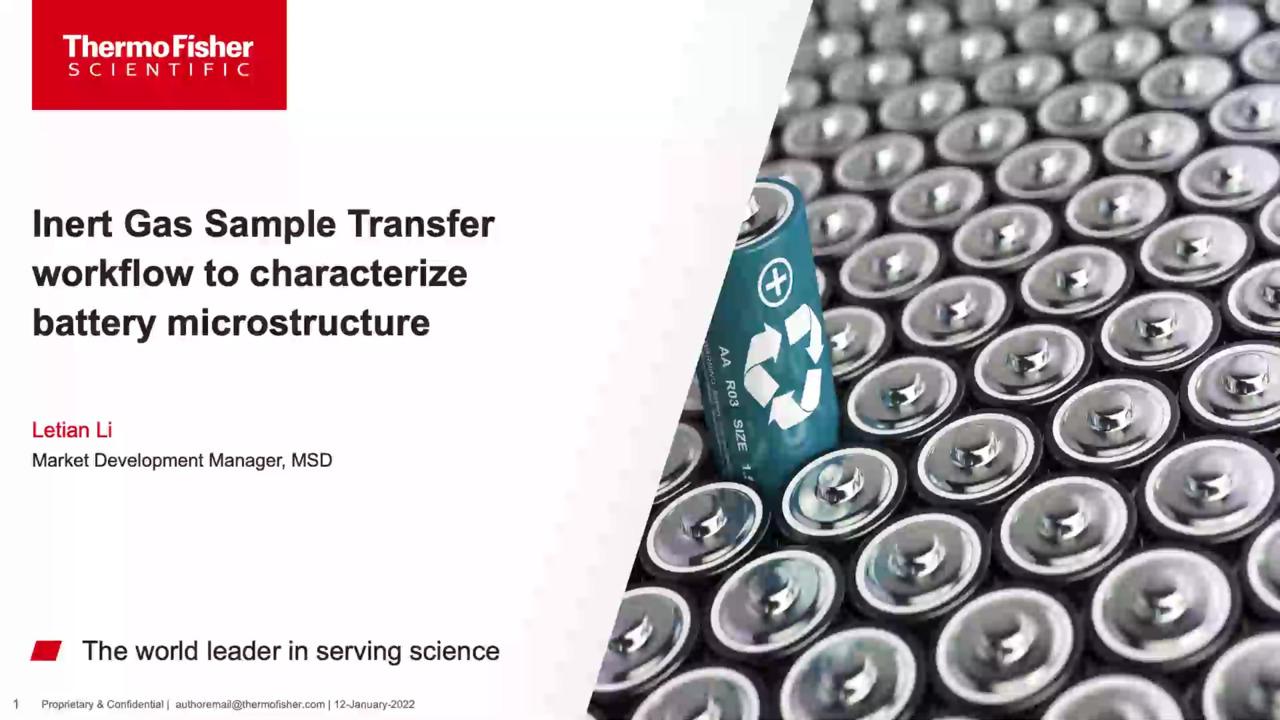
Avoid contamination of air-, moisture-, and/or beam-sensitive battery samples during preparation and sample transfer |
IGST workflow: DualBeam, SEM/Desktop SEM (in glove box), TEM, Avizo, CleanConnect |
Complete workflow to enable sample characterization of sensitive battery materials in their native state without contamination |
|
Detection of lithium is difficult using SEM, EDS, and TEM |
TOF-SIMS |
Accurately detect and map lithium in battery samples in 2D and 3D down to 10 ppm |
App note: Ion spectroscopy using TOF-SIMS on a Thermo Scientific Helios DualBeam |
TEM |
iDPC technology can clearly image light elements like lithium at atomic scale |
App note: Integrated Differential Phase Contrast on Talos S/TEM |
|
Characterize battery structure at different scales beyond the capacity of a single instrument |
CT, SEM, Raman, DualBeam, Avizo, EDS |
Correlative workflow allowing multiscale imaging and analysis of battery microstructure |
App note: Multiscale image-based control and characterization of lithium-ion batteries |
| App note: Multiscale 3D imaging solutions for Li-ion batteries | |||
Prepare a large 2D area on the sample surface with high polishing quality for 2D imaging and characterization |
DualBeam (Plasma FIB-SEM), EDS |
High-throughput automated spin mill with high surface quality |
App note: Large area automated sample preparation for batteries |
SEM, CleanMill |
CleanMill offers a dedicated workflow for air-sensitive samples, an ultra-high energy ion gun for fast polishing, and a cryogenic function to protect sample integrity |
||
Characterize key microstructure properties (like tortuosity) for electrode structure performance correlations |
DualBeam, EDS, TOF-SIMS, Avizo |
3D characterization of battery structure · Hardware to image 3D battery structure at different scales · Software to automate 3D imaging data collection · Thermo Scientific Avizo Software workflow for image analysis and quantification |
Blog post/video: Advancing lithium-ion battery technology with 3D imaging |
App note: Multiscale image-based control and characterization of lithium-ion batteries |
|||
Characterize beam-sensitive materials like SEI at nanoscale |
TEM, EDS, Avizo |
Nano- and atomic-scale characterization of energy materials · Cryo-EM workflow for accurate data collection with superior EDS performance · Avizo Software for structure quantification and visualization |
Brochure: Analytical solutions for battery and energy storage technology |
In situ kinetic analysis (like heating) via electron microscope |
SEM |
Multiple in situ heating stage choices with integrated software for Thermo Scientific SEMs to understand cathode synthesis mechanisms |
Brochure: Scanning electron microscopy for lithium battery research |
Characterize beam-sensitive separator samples without damage |
SEM/SDB |
Superior low-KeV imaging and a cryo-FIB milling solution allow characterization of separator microstructure in 2D and 3D |
App note: Strategies for accurate imaging on battery separator structure |
Probe intrinsic SEI within a coin cell via electron microscopy |
Laser Plasma FIB |
High-energy, high-milling rate laser enables direct cross-section milling to understand Li-metal cell degradation mechanism |
|
Understand stoichiometry of solid electrolyte film as a function of depth |
XPS |
XPS depth profiling can quantify elements at each depth |
|
Measure electrode surface chemistry |
XPS |
XPS can quantify the chemical states present at the electrode surface |
App note: Analysis of electrode materials for lithium ion batteries |
Track the evolution of the SEI layer |
XPS |
Materials can be depth profiled using XPS and a cluster ion source to follow the development of the SEI layer after cycling |
|
In situ electrode cycling |
XPS |
Electrodes can be operated in situ to monitor spectral changes as they are charged and discharged |
|
Investigate changes in separator chemistry during cell lifetime |
XPS |
The surface chemistry of polymeric materials is easily characterized using XPS |
|
Profile battery components ex situ without missing point-to-point variability across an area |
Raman |
Raman microscopy can be used to look at changes to materials and distributions of components that occurred during use or testing |
|
Identify phases and determine structures in anodes and cathodes |
Raman |
Raman microscopy can visually show the spatial distribution of different phases of the same material with different performance characteristics |
App note: Raman analysis of lithium-ion batteries – Part I: Cathodes |
App note: Raman analysis of lithium-ion batteries – Part II: Anodes |
|||
XRD |
XRD can help to identify and quantify specific polymorphic structures of interest to increase yield and efficiency |
Brochure: ARL EQUINOX 100 X-ray Diffractometers | |
Trace and map anode composition across charge and discharge cycles |
Raman |
Raman microscopy can be used for in situ monitoring of changes on electrode surfaces during charge/discharge cycles |
|
Confirm the presence of specific carbon allotropes as anode components and in hybrid materials |
Raman |
Raman spectroscopy is particularly adept at the analysis of allotropes of carbon, including carbon in hybrid materials |
App note: Raman analysis of lithium-ion batteries – Part II: Anodes |
Understand the association of ionic species and distribution of components in solid polymer electrolytes (SPEs) |
Raman |
Raman microscopy can be used to visualize the spatial distribution of components in SPEs and indicate ionic associations |
App note: Raman analysis of lithium-ion batteries – Part III: Electrolytes |
Study crystallinity, stability, and reactivity in battery materials |
XRD |
X-ray diffraction can determine the percentage of crystallinity vs amorphous content of the active material, as well as structural stability and repeatability in real time |
|
Follow charge/discharge reactions in situ |
XRD |
During charge/discharge, the cathode and anode of every battery cell undergo changes. XRD allows you to follow the changing phase composition and the evolution of the crystalline structure |
Abbreviations: Avizo = Avizo Software; CT = Computed tomography; DualBeam = Focused ion beam scanning electron microscopy (FIB-SEM); EDS = Energy-dispersive X-ray spectroscopy; FIB = Focused ion beam; FTIR = Fourier transform infrared spectroscopy; iDPC = Integrated differential phase contrast; IGST = Inert sample gas transfer; SDB = Small DualBeam; SEI = Solid electrolyte interface; SEM = Scanning electron microscopy; SPE = Solid polymer electrolytes; TEM = Transmission electron microscopy; TOF-SIMS = Time of flight secondary ion mass spectrometry; XPS = X-ray photoelectron spectroscopy; XRD = X-ray diffraction.
For Research Use Only. Not for use in diagnostic procedures.