Search Thermo Fisher Scientific
Volumescope 2 Scanning Electron Microscope for serial block face imaging
The Thermo Scientific Volumescope 2 Scanning Electron Microscope (SEM) is our state-of-the-art serial block-face imaging system. Keep control of experiments with easy to use technology and protect a wide range of possible samples with tried and tested solutions for every acquisition step.
Block face imaging
Unraveling the complex 3D architecture of cells and tissues in their natural context is crucial for structure-function correlation in biological systems and soft materials. Serial block-face SEM (SBF-SEM) combines in situ sectioning and imaging of plastic-embedded tissue blocks within the SEM vacuum chamber for automated, 3D reconstruction of large tissue volumes. Axial resolution was once limited by the minimum section thickness, but truly isotropic resolution is now possible with the addition of multi-energy deconvolution.
3D SEM
Following in situ sectioning of the block-face using a diamond knife, the freshly exposed tissue is imaged several times using increasing accelerating voltages. These images are subsequently used in a deconvolution algorithm to derive several optical subsurface layers, forming a 3D subset. By repeating this cycle, the Volumescope 2 SEM offers isotropic datasets with 10 nm Z-resolution.


Polymer microstructures
High-resolution imaging in 3D is of great importance when trying to understand the microstructure and properties of polymer materials. The Volumescope 2 SEM combines in situ sectioning and imaging of polymers within the SEM vacuum chamber, in a fully automated fashion, for reconstruction of large volumes with truly isotropic 3D resolution. Visualizing a large volume at such resolutions is critical for revealing how regions with different microstructures affect the overall properties of the material. For example, the Volumescope 2 SEM can recreate a large representative volume of a filtration membrane, providing accurate transport properties that lead to enhanced performance predictions for new filter designs.

Easy to use technology
Keep control of experiments with easy to use technology. Reuse jobs and system settings and select multiple regions of interest during job acquisition.
Visualize and navigate during acquisition
Visualize and navigate during acquisition with Thermo Scientific Amira Live Tracker Software to optimize/control your outcome/Automate large 3D volume acquisitions as well as reconstructions.
Protect valuable samples
Protect valuable samples with tested solutions at every acquisition step: debris trap and swipe features ensure sample quality; low vacuum detector enables imaging of highly charged samples.
Easy and fast mounting microtome exchange
Easy and fast mounting microtome exchange for normal SEM operation or automated array tomography with the addition of Thermo Scientific Maps Software.
| Electron optics |
|
| Source lifetime | 24 months |
| Automation |
|
| Stage | Double stage scanning deflection |
| Electron beam |
|
| Chamber |
|
| Detectors |
|
| Vacuum system |
|
| Sample holders |
|
See datasheet for full list of specifications including software, accessories and a summary of installation requirements.

Investigación sobre patologías
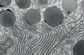
La microscopía electrónica de transmisión (TEM) se utiliza cuando la naturaleza de la enfermedad no puede establecerse mediante métodos alternativos. Con las imágenes nano-biológicas, TEM proporciona información precisa y fiable para ciertas patologías.

Investigación sobre biología de plantas
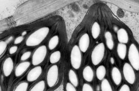
La investigación fundamental de la biología de las plantas se hace posible mediante la criomicroscopía electrónica, que proporciona información sobre proteínas (con análisis de partículas individuales), a su contexto celular (con tomografía), hasta la estructura general de la planta (análisis de grandes volúmenes).

Investigación sobre materiales fundamentales
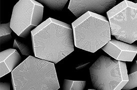
Se investigan nuevos materiales a escalas cada vez más pequeñas para lograr el máximo control de sus propiedades físicas y químicas. La microscopía electrónica proporciona a los investigadores información clave sobre una amplia variedad de características materiales a escala nanométrica.

Control de calidad
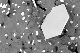
El control y garantía de calidad son esenciales en la industria moderna. Ofrecemos una gama de herramientas de EM y espectroscopía para el análisis multiescala y multimodal de defectos, lo que le permite tomar decisiones fiables e informadas para el control y la mejora de procesos.

Análisis de grandes volúmenes
Una novedosa solución de adquisición de imágenes de cara de bloques en serie (SBFI) que combina la microscopía electrónica de barrido de deconvolución de la multienergía (MED-SEM) con cortes en secciones in situ. La automatización y las funciones fáciles de utilizar proporcionan una solución isotrópica para muestras de grandes volúmenes.
Servicios de microscopía electrónica
Para garantizar un rendimiento óptimo del sistema, le proporcionamos acceso a una red de expertos de primer nivel en servicios de campo, asistencia técnica y piezas de repuesto certificadas.


