Search Thermo Fisher Scientific

The standard configuration for the Thermo Scientific Tundra Cryo-TEM includes the Thermo Scientific Ceta F CMOS Camera, which is equipped with a dose-fractionation mode and offloading functionality for high-resolution protein reconstruction. Acquisition of dose-fractions can reduce the sample drift and motion caused by beam induced movement. The Tundra Cryo-TEM can also be configurated with the Thermo Scientific Falcon C Direct Electron Detector, designed specifically for this platform. It provides higher resolution capabilities than the Ceta F CMOS Camera, with a unique combination of high image quality, high throughput, and efficient lossless data compression through electron-event representation (EER).
Falcon C Direct Electron Detector
The Falcon C Direct Electron Detector, designed for the Tundra Cryo-TEM, enables you to obtain high-resolution structures to reveal new biological insights at 100 kV. The Falcon C DED provides higher resolution capabilities than the Ceta F CMOS Detector, offering a unique combination of high image quality, high throughput, and efficient lossless data compression enabled via electron-event representation (EER), providing a powerful productivity and performance boost.
Falcon C Direct Electron Detector Features

Falcon C Direct Electron Detector Specifications
Camera architecture |
Direct electron detection |
Sensor size |
2,048 x 2,048 pixels, 5.7 x 5.7 cm2 |
Pixel size |
28 x 28 μm2 |
TEM Operating voltage |
100 kV |
Internal frame rate |
250 fps |
Frame rate to storage |
240 fps (EER mode) |
Camera overhead time |
0.5 s per acquisition |
File formats |
EER (native), MRC as integrated image |
Detection modes |
Electron counting mode Survey mode (fast linear mode) |
Automation |
Integrated in Tundra software (including EPU) |
Imaging performance in EER mode1 |
100kV |
DQE(0) DQE(½ Nyquist) DQE(1 Nyquist) |
≥ 0.95 ≥ 0.55 ≥ 0.2 |
Ceta-F CMOS Camera
The Ceta-F Camera is a scintillator-based camera optimized for low-dose imaging for sensitive samples. It is ideal for working with dose-sensitive materials, such as proteins embedded in vitreous ice. The camera’s large field of view (4k x 4k), in combination with integration mode, allows fast sample screening and data collection. To better facilitate cryo-EM applications, dose fractions are stored, which allows further image quality improvement during the 3D reconstruction. Optimizations, such as motion correction and radiation damage compensation, can be applied to the fractions. High-resolution 3D reconstructions, especially for large (>200kDa) and symmetric protein complexes, are possible.
* non-AFIS mode
Aberration-free image shift
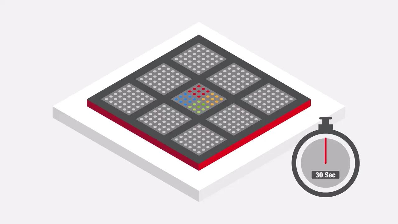
Aberration-free image shift (AFIS) functionality uses image beam shifts, which are instant and stable. AFIS saves time by eliminating the need to perform mechanical stage movement and stabilization. AFIS accelerates the data collection 4x, from approximately 80 images/hour to 320 images/hour. Both the Falcon C and Ceta-F CMOS detectors for the Tundra Cryo-TEM work with AFIS mode.
Cryo-EM Software
Our cutting-edge cryo-electron microscopy software enhances the capabilities of this powerful imaging technique, helping you achieve insights into the structure and function of a wide range of targets.
Smart EPU Software
The Tundra Cryo-TEM comes with a complete suite of automation software for the efficient optimization of your sample’s biochemistry and determination of its structure. This includes user-friendly single particle data acquisition software, Thermo Scientific EPU Software, which offers guided day-to-day operation, a traffic light UI element that indicates the microscope’s status, and predefined templates for typical use cases that allow you to begin collecting high-resolution data with only a few clicks. Smart EPU Software, an AI-enabled software solution, is capable of analyzing intermediate results, providing instant feedback, and steering data collection on the fly. The AI algorithms are based on years of cryo-EM knowledge, designed to replace decisions that experts need to make upfront and ensure that your instrument is working at optimal conditions, allowing you to focus on the science rather than on fine-tuning the microscope.
EPU Software user interface
The embedded traffic light UI element indicates the microscope’s status. Predefined templates for typical use cases allow you to begin collecting high-resolution data with only a few clicks.
AI-enabled software
Smart EPU Software’s automation and AI-driven plugins guide the user through critical decision points and, in some cases, make decisions automatically, reducing the need for manual user intervention.
For Research Use Only. Not for use in diagnostic procedures.
