Search Thermo Fisher Scientific
Mounting Coverslips Protocol

Formulations of mounting media that can add favorable properties such as optimizing the refractive index to match that of glass, preventing photobleaching, or preserving samples for long-term storage are widely available. Keep in mind that hard-setting mountants require time to “cure” or harden.
What you need
- Fixed, permeabilized, blocked, and stained sample on coverslip in a saline-based buffer solution such as PBS
- Glass microscope slide
- Mounting medium
Mounting coverslips protocol
Try to limit the formation of bubbles in your mounting medium. Some bubbles are inevitable, and you can image around them as long as there are only a few.
To avoid bubbles:
- Don’t shake or invert your bottle of mounting medium.
- Clear bubbles from the tip of your applicator (i.e., from the tip of the pipette or dropper bottle) by squeezing a little bit of mounting medium onto a lab tissue before applying mounting medium to your slide
- Clean the surface of the non-sample side of the coverslip prior to imaging. Fingerprints, residual salts, and other contaminants can diminish image quality.
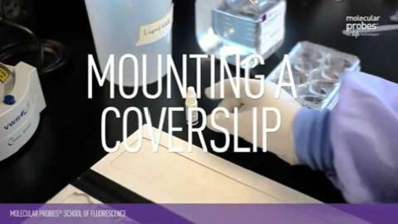
Mounting a Coverslip
This video explains step-by-step how to mount a coverslip properly for cellular imaging as illustrated in steps 1-4 of the below protocol.
| 1 |  | Apply a small amount of mounting medium to the surface of the slide; try to use an amount that will just fill the space under the coverslip. |
| 2 |  | Remove coverslip containing the sample from the buffer. |
| 3 |  | Blot excess buffer or solvent from the non-sample surface of the coverslip (or allow it to air-dry and then remove the salt residue before imaging). |
| 4 |  | Slowly tip the coverslip onto the mounting medium, and avoid creating bubbles as you lower it into place. |
| 5 |  | Follow manufacturer’s directions for curing time |
| 6 |  | Optional: Seal the coverslip if desired. |
| 7 |  | Image. |
Share
For Research Use Only. Not for use in diagnostic procedures.