Search Thermo Fisher Scientific
Nuclear Apoptosis Assays for Flow Cytometry

Various stages of apoptosis are characterized by changes in nuclear morphology, including DNA fragmentation, chromatin condensation, degradation of nuclear envelope, nuclear blebbing, and DNA strand breaks. We offer a variety of unique reagents for analyzing nuclear changes during apoptosis; many are compatible with reagents used to detect additional apoptotic parameters using flow cytometry.
Nuclear chromatin condensation
Cells undergoing apoptosis display an increase in nuclear chromatin condensation. As the chromatin condenses, cell-permeable nucleic acid stains becomes hyperfluorescent, thus enabling the identification of apoptotic cells when combined with a traditional dead-cell stain.

Figure 1. Analysis of chromatin condensation with Vybrant DyeCycle Violet stain and SYTOX AADvanced stain. Jurkat cells (T-cell leukemia, human) were treated with 10 µM camptothecin for 6 hours (3B) or left untreated as a control (3A). Cells were then mixed with the reagents in the Apoptosis Kit- Vybrant DyeCycle Violet/SYTOX AADvanced Kit and analyzed by flow cytometry using 405/488 nm dual excitation. Note that the campthothecin-treated cells (3B) have a higher percentage of apoptotic cells than the basal level of apoptosis seen in the control cells (3A). A=apoptotic cells, V = viable cells, N = necrotic cells.
| Product | Laser | Ex/Em | Cat. No. |
|---|---|---|---|
| Hoechst 33342 | UV | 350/461 | H1399 |
| Vybrant DyeCycle Violet | 405 | 369/437 | V35003 |
| YO-PRO-1 iodide | 488 | 491/509 | Y3603 |
| TO-PRO3 Ready Flow Reagent | 633 | 642/661 | R37170 |
| Condensed chromatin stain | Dead cell stain | Lasers | Ex/Em | Cat. No. |
|---|---|---|---|---|
| Hoechst 33342 | PI | UV and 488 nm | Hoechst 33342: 350/461 nm PI: 535/617 nm | V13244 |
| YO-PRO-1 Hoechst 33342 | PI | UV and 488 nm | Hoechst 33342: 350/461 nm YO-PRO-1: 491/509 nm PI: 535/617 nm | V23201 |
| Vybrant DyeCycle Violet stain | SYTOX AADvanced | 405 and 488 nm | Vybrant DyeCycle Violet stain: 369/437 nm SYTOX AADvanced: 546/647 nm | A35135 |
| YO-PRO-1 | PI | 488 nm | YO-PRO-1: 491/509 nm PI: 535/617 nm | V13243 |
| PO-PRO-1 | 7-AAD | UV and 488 nm | PO-PRO-1: 434/456 nm 7-AAD: 546/647 nm | V35123 |
DNA fragmentation (TUNEL assay)
DNA fragmentation that occurs during apoptosis produces DNA strand breaks, and can be analyzed using TUNEL (terminal deoxynucleotidyl transferase dUTP nick end labeling) assays. The APO-BrdU TUNEL assay is a two-color assay for labeling DNA breaks and total cellular DNA to detect apoptotic cells by imaging or flow cytometry. Propidium iodide is also included to analyze total DNA content or cell-cycle phase.
| Product | Laser | Ex/Em | Cat. No. |
|---|---|---|---|
| APO-BrdU TUNEL Assay Kit with Alexa Fluor 488 anti-BrdU | 488 | 495/519 | A23210 |
Resources
Fluorophore and reagent selection guide for flow cytometry
Download Flow Cytometry Protocols Handbook
Spectral Flow Cytometry Fundamentals
Invitrogen eBioscience Resources—Selection guides, Best Protocols, product performance and more.
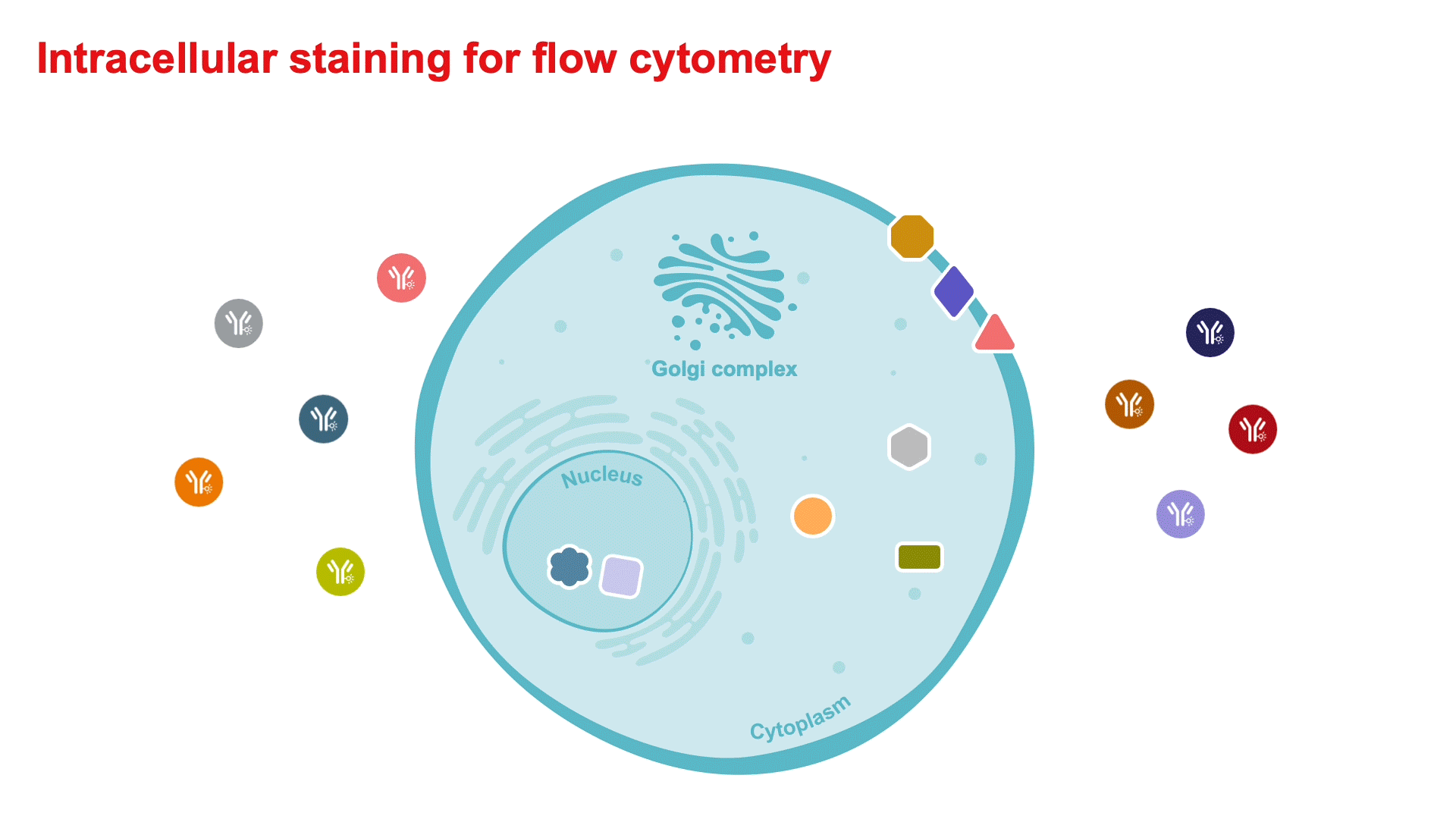
Intracellular Staining for Flow Cytometry How-To Video—for detecting cytokines and intranuclear markers.
Flow Cytometry Learning Center—Access flow cytometry educational resources for better experiment planning and execution.
Flow Cytometry Panel Builder—Design your flow cytometry panel with this online tool for a simplified, customizable experience to fit your needs.
5 Steps Resources
Support
Flow Cytometry Support Center—Find technical support recommendations for your flow cytometry workflows, including tips for experimental setup and in-depth troubleshooting help.
Flow Cytometry Panel Design Support—Work with one of our technical sales specialists to discuss your experimental needs and guide you through the process.
Not for resale. Super Bright Polymer Dyes are sold under license from Becton, Dickinson and Company.
For Research Use Only. Not for use in diagnostic procedures.