Search Thermo Fisher Scientific
FIX & PERM Cell Fixation & Cell Permeabilization Kit

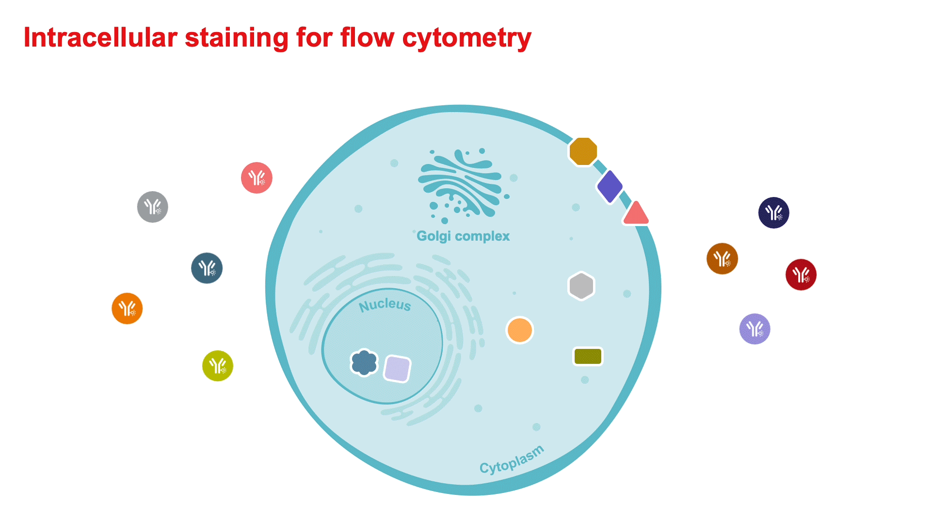
The FIX & PERM Cell Fixation and Cell Permeabilization Kit achieves mild fixation and permeabilization of cells that leaves their morphological scatter characteristics intact, enabling researchers to accurately identify previously undetectable intracellular markers, such as cytoplasmic or nuclear enzymes, oncoproteins, cytokines, and immunoglobulins.
View all FIX & PERM cell fixation & permeabilization products
On this page
- Compatible with analysis of most cellular antigens
- No effect on cellular morphological scatter
- Reduced background staining
- Proven protocols
The FIX & PERM Cell Permeabilization Kit consists of matched Fixation Reagent (Medium A) and Permeabilization Reagent (Medium B) for simultaneous analysis of intracellular and cell-surface antigens in the same cell population (Figure 1). This procedure facilitates antibody access to intracellular structures and leaves the morphological light-scattering characteristics of the cells intact. These formulations reduce background staining and allow simultaneous addition of permeabilization medium and fluorophore-labeled antibodies. The Fixation Medium and Permeabilization Medium are available separately as well.
FIX & PERM reagents are suitable for the analysis by flow cytometry of normal and malignant leukocyte populations derived from various human biological samples (blood, bone marrow, and others) (Figure 2).

Figure 1. Use of FIX & PERM Cell Permeabilization Kit for simultaneous surface antigen and intracellular antigen staining. C57BL/6 splenocytes were left unstimulated or stimulated for 5 hours with phorbol myristate acetate (PMA) and ionomycin in the presence of brefeldin A. Cells were then surface-stained with fluorescein (FITC)-conjugated anti–mouse CD4 antibody. This step was followed by fixation and permeabilization of the sample using the FIX & PERM Cell Permeabilization Kit. Intracellular staining was performed during the permeabilization step using PE-Cy7 tandem–conjugated anti–mouse γ-interferon (γ-IFN) antibody and Pacific Blue dye–conjugated anti–mouse tumor necrosis factor α (TNF-α) antibody. Data were collected using the Attune Flow Cytometer (blue/violet) with 488 nm excitation and a 530/30 nm bandpass emission filter to detect FITC fluorescence and a 640 nm longpass filter to detect PE-Cy7 tandem fluorescence. Pacific Blue conjugate fluorescence was detected using 405 nm excitation and a 450/40 nm bandpass emission filter. (Top row) γ-IFN and TNF-α antibody co-staining of total mouse splenocytes, gated on lymphocytes, that were left unstimulated (left) or stimulated (right) with PMA and ionomycin in the presence of brefeldin A. (Bottom row) CD4+ T cell expression of TNF-α (left) and γ-IFN (right) after stimulation as described above.

Figure 2. Immunophenotyping of a whole-blood sample. Two-day-old whole blood was lysed with ammonium chloride. Cells were first stained with CD13-APC conjugate. Cells were washed, stained with LIVE/DEAD Fixable Near-IR dead cell stain, washed, and fixed with FIX & PERM Reagent A. Cells were then washed, permeabilized with FIX & PERM Reagent B, stained with MPO-FITC conjugate, and washed. Cells were analyzed on a BD™ LSRII flow cytometer. Gating on live cells (generally recommended, to eliminate dead cells) was performed. Further subgating is recommended in order to obtain the most accurate results.
The FIX & PERM Cell Fixation & Permeabilization Kit allows efficient detection of a wide variety of markers and intracellular proteins, including, but not limited to:
| Lysosomal proteins | Elastase, lactoferrin, lysozyme, myeloperoxidase, proteinase-3 |
| Cytoplasmic CD molecules | CD3, CD13, CD22, CD62P, CD63, CD68, CD79a |
| Nuclear proliferation markers | BrdU, Ki-67, PCNA, nuclear enzymes, TdT |
| Oncoproteins | Bcl-2, c-Myc, p53, HIV antigens, p24 |
| Cytokines & chemokines (human) | IFN-γ, TNF-α, IL1-β, IL-2, IL-4, IL-10, IL-12, IL-13, IL-16, RANTES |
| Cytokines & chemokines (mouse) | IFN-γ, TNF-α, IL-2, IL-4, IL-5, IL-10, IL-12, IL-13, IL-16 |
| Immunoglobulins (human) | IgA, IgG, IgD, IgM, kappa, lambda |
| Immunoglobulins (mouse) | IgG, IgM |
| Other molecules | ZAP-70 (zeta-associated protein 70), MHC class II, phosphotyrosine, thrombospondin, cyclins, transfected cells, MDR (multidrug resistance) |
FIX & PERM cell fixation & permeabilization reagents are designed for use with all commercially available flow cytometers. This standard procedure for intracellular staining with FIX & PERM fixation & permeabilization reagents gives optimal results for most antigens. A modification using precooled absolute methanol has been shown to give better results for certain cell cycle antigens such as Ki-67 and PCNA when using FITC-conjugated antibodies. The methanol step is not recommended when using RPE-conjugated antibodies. These staining protocols are intended for use directly with the cell suspension to be analyzed.
Other lysing solutions should not be used prior to the use of FIX & PERM cell fixation & permeabilization reagents.
- For each sample to be analyzed, add the appropriate volume of the conjugated antibody directed to the cell surface marker(s) of interest, and/or the appropriate isotype control(s), to an appropriate 5 mL, 12 x 75 mm tube.
- Pipette 100 μL of whole blood or adjusted cells (stimulated PBMCs, bone marrow, spleen, thymus), equivalent to 1 x 10⁶ cells, into each tube containing the conjugated antibody or isotype control.
- Vortex each tube gently to mix, and incubate for 15 minutes in the dark at room temperature.
- Add 100 μL of Reagent A (Fixation Medium) and incubate for 15 minutes in the dark at room temperature.
- Wash once with 3 mL wash medium (PBS + 0.1% NaN3 + 5% FBS).
- Centrifuge for 5 minutes at 300–350 x g, aspirate supernatant, and vortex to fully resuspend the cell pellet.
- Add 100 μL of Reagent B (Permeabilization Medium) and the recommended volume of intracellular antibodies or the corresponding isotype control(s).
- Vortex 1–2 seconds and incubate for 20 minutes in the dark at room temperature.
- Wash once with 3 mL wash medium (PBS + 0.1% NaN3 + 5% FBS).
- Centrifuge for 5 minutes at 300–350 x g and aspirate the supernatant.
- Resuspend cells in sheath fluid for immediate analysis, or fix in 0.5 mL of 0.1% paraformaldehyde and store at 2–8°C in the dark. Fixed cells should be analyzed within 18 hours.
The methanol modification procedure is recommended for cell cycle antigens such as BrdU, Ki-67, and PCNA when using FITC-conjugated antibodies. It is not recommended when using RPE-conjugated antibodies.
- For each sample to be analyzed, add 100 μL of adjusted cell volume (equivalent to 1 x 10⁶ cells) to an appropriate 5 mL, 12 x 75 mm tube.
- Add 100 μL of Reagent A (Fixation Medium) and incubate for 2–3 minutes at room temperature.
- Add 4 mL of precooled (0–4°C) absolute methanol and vortex.
- Incubate for an additional 10 minutes at 0–4°C.
- Centrifuge for 5 minutes at 300–350 x g and wash with wash medium (PBS + 0.1% NaN3 + 5% FBS).
- Add 100 μL of Reagent B (Permeabilization Medium) and an appropriate volume of intracellular antibodies or the corresponding isotype control(s).
- Vortex at low speed for 1–2 seconds and incubate for 30 minutes at room temperature.
- Wash once with 3 mL wash medium (PBS + 0.1% NaN3 + 5% FBS).
- Centrifuge for 5 minutes at 300–350 x g and aspirate the supernatant.
- Resuspend cells in sheath fluid for immediate analysis, or fix in 0.5 mL of 0.1% paraformaldehyde and store at 2–8°C in the dark. Fixed cells should be analyzed within 18 hours.
VERY IMPORTANT: Blood must be collected into heparinized tubes.
- Dispense 0.5 mL of heparinized whole blood into each of two 12 x 75 mm snap-cap tubes.
- Add 0.5 mL of RPMl 1640 (without additives) to each tube to bring the volume to 1 mL.
- To the first tube, add 10 µg brefeldin A (20 µL of a stock solution of brefeldin A at a concentration of 0.5 mg/mL in 100% EtOH and stored at –20°C). Mix contents of the tube gently. This tube represents the resting cell population.
- To the second tube, add 10 µg brefeldin A, 25 ng PMA (2.5 µL of a stock solution of PMA at a concentration of 1 mg/mL in DMSO and diluted 1:100 in RPMI 1640 (without additives) and stored at –20°C), and 1 µg ionomycin (1 µL of a stock solution of ionomycin at a concentration of 1 mg/mL in DMSO and stored at –20°C). Mix contents of the tube gently. This tube represents the activated cell population.
- Incubate for 4 hours at 37°C in a 7.5% humidified CO2 incubator.
- At the end of the incubation period, mix the cells again and aliquot 100 µL into 12 x 75 mm snap-cap tubes.
- Add 5 µL of the appropriate antibodies for phenotyping cells (cell-surface staining) (e.g., CD3-TRI-COLOR, CD4-TC, CD8-TC, CD45-TC, or CD69-TC for a three-color staining assay; either FITC or RPE may be appropriate for a two-color assay).
- Incubate in the dark for 15 minutes at room temperature.
- Add 100 µL of Reagent A from the FIX & PERM Kit. Mix cells gently and incubate for an additional 15 minutes at room temperature in the dark.
- Wash twice with wash medium (PBS + 1 % BSA, 0.1 % NaN3, 1 % FBS). The pellet becomes less cohesive on the second wash, so use caution to avoid losing cells.
- To the washed cells add 100 µL of Reagent B from the FIX & PERM Kit and 1–10 µL of each anti-cytokine antibody OR isotype control.
- Incubate for 20 minutes at room temperature.
- Wash twice with wash medium and resuspend pellet in FACS fix.
- Analyze by gating on SSC and FL-3 (3- or 4-color assay).
- Stahl M et al. (2009) CEA in activated macrophages. New diagnostic possibilities for tumor markers in early colorectal cancer. Anticancer Res 29(8):3245–3251. (Notch)
- Vang KB et al. (2008) Roles of Pofut1 and O-Fucose in Mammalian Notch Signaling. J Biol Chem 283:13638–13651. (phosphor-STAT5)
- LeibundGut-Landmann S et al. (2008) Stimulation of dendritic cells via the dectin-1/Syk pathway allows priming of cytotoxic T cell responses. Blood 112(13): 4971–4980.
- Rosen HR et al. (2008) Pretransplantation CD56+ innate lymphocyte populations associated with severity of hepatitis C virus recurrence. Liver Transpl 14:31–40. (IFNy)
- Basu S et al. (2008) Foxp3-mediated induction of Pim 2 allows human T regulatory cells to preferentially expand in rapamycin. J Immunol 180:5794–5798. (IL-2)
- Libani IV (2008) Decreased differentiation of erythroid cells exacerbates ineffective erythropoiesis in β-thalassemia. Blood 112(3): 875-885. (phospho-JAK2)
- El Shikh MEM et al. (2009) T-independent antibody responses to T-dependent antigens: a novel follicular dendritic cell-dependent activity. J Immunol 182: 3482-3491. (Phosphotyrosine)
- Nieto WG et al. (2009) Increased frequency (12%) of circulating chronic lymphocytic leukemia–like B cell clones in healthy subjects using a highly sensitive multicolor flow cytometry approach. Blood 114(1):33–37.
- Meyer zum Büschenfelde C et al. (2010) Recruitment of PKC-βII to lipid rafts mediates apoptosis-resistance in chronic lymphocytic leukemia expressing ZAP-70. Leukemia 24:141–152.
Resources
Fluorophore and reagent selection guide for flow cytometry
Download Flow Cytometry Protocols Handbook
Spectral Flow Cytometry Fundamentals
Invitrogen eBioscience Resources—Selection guides, Best Protocols, product performance and more.
Intracellular Staining for Flow Cytometry How-To Video—for detecting cytokines and intranuclear markers.
Flow Cytometry Learning Center—Access flow cytometry educational resources for better experiment planning and execution.
Flow Cytometry Panel Builder—Design your flow cytometry panel with this online tool for a simplified, customizable experience to fit your needs.
5 Steps Resources
Support
Flow Cytometry Support Center—Find technical support recommendations for your flow cytometry workflows, including tips for experimental setup and in-depth troubleshooting help.
Flow Cytometry Panel Design Support—Work with one of our technical sales specialists to discuss your experimental needs and guide you through the process.