Search Thermo Fisher Scientific
CellEvent Senescence Green Probe for fluorescence imaging and flow cytometry
(See a list of the products featured in this article.)
Cell biologists are focusing on the causes and treatments of age-related diseases, given increased life expectancies and an aging human population. Cellular senescence—exhibited by cells that have stopped dividing but remain metabolically active—is an important pathway for controlling unlimited cell division. However, if senescent cells are not removed by the immune system, a chronic pro-inflammatory environment ensues that can affect nearby healthy cells and increase the risk of age-related diseases. Senescent cells are more prevalent with increased age and are thought to play a role in many age-related pathologies, including neurodegenerative diseases such as Parkinson’s and Alzheimer’s as well as inflammatory diseases such as osteoarthritis and cardiovascular disease [1].
Recent studies in mouse models have shown that specific targeting of senescent cells with a senolytic cocktail produced longer healthspans and lifespans [2]. The development of senolytic drugs, which can selectively kill senescent cells or inhibit cellular senescence, is dependent on characterization of the senescence pathway and its signaling molecules, as well as on development of research tools for identifying senescent cells in various tissues and biological samples.
Multifactor detection of senescence
Senescent cells are characterized by the release of pro-inflammatory cytokines and chemokines, increased beta-galactosidase activity (senescence-associated (SA) β-Gal), heterochromatin foci (SAHF), and changes in cellular morphology [3]. Because there is no single marker of senescence, the identification of senescent cells requires correlating the presence of multiple biomarkers, including proteins involved in cell cycle arrest (e.g., p16 and p21), proteins associated with DNA damage (e.g., pH2AX), and increased β-Gal expression.
The detection of senescence based on the upregulation of β-Gal can be accomplished using a β-Gal substrate such as X-Gal (5-bromo-4-chloro-3-indolyl β-D-galactopyranoside), a colorimetric substrate that produces a blue-green precipitate upon enzymatic cleavage. Since the mid-1990s, X-Gal has been considered the gold standard for labeling senescent cells in tissue and biological samples [4]; yet, the X-Gal assay has several drawbacks, including its long incubation time and the difficultly of combining it with other staining protocols to identify other senescence biomarkers, which limits its usefulness in the development of senolytic therapies. Additionally, colorimetric X-Gal staining is only useful for brightfield microscopy applications; it cannot be combined with fluorescence immunostaining or detected by flow cytometry. The fluorogenic substrate C12FDG (5-dodecanoylaminofluorescein di-β-D-galactopyranoside) has also been used for β-Gal detection; however, its fluorescent product is prone to leakage from the cell, making it incompatible with immunostaining protocols.
CellEvent Senescence Green: A fluorogenic senescence-associated β-Gal probe
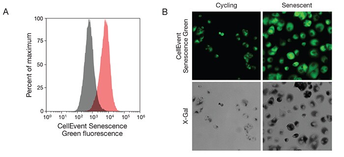
To address the limitations of current senescence detection methods, we have developed the Invitrogen CellEvent Senescence Green Probe. This fluorogenic β-Gal substrate is designed for the detection of senescent cells by fluorescence imaging or flow cytometry, based on the upregulation of SA β-Gal (Figure 1). The CellEvent Senescence Green Probe contains two galactoside moieties, as well as an additional moiety that reacts with several functional groups found in proteins. This nonfluorescent substrate is cleaved by intracellular β-Gal to produce a bright green-fluorescent product (Ex/Em = 490/514 nm) that is well retained in cells due to its covalent binding to intracellular proteins. In addition, the CellEvent Senescence Green Probe is easy to use: simply fix the cells, add the reagent, incubate, and detect fluorescence by imaging or flow cytometry.

Figure 1. Detection of senescent cells with CellEvent Senescence Green Probe by flow cytometry and fluorescence imaging. T47D cells were treated with 5 μM palbociclib for 16 days or left untreated. After treatment, cells were fixed with 4% formaldehyde for 10 min at room temperature, followed by senescence staining for 90 min in a 37ºC incubator with no CO2. (A) Flow cytometric analysis was performed using the Invitrogen CellEvent Senescence Green Flow Cytometry Assay Kit on the Invitrogen Attune NxT Flow Cytometer, with 488 nm excitation and a 530/30 nm emission filter. The histogram shows clear separation of treated senescent cells (pink) from untreated cycling cells (gray). (B) Imaging analysis was performed using the Invitrogen CellEvent Senescence Green Detection Kit on the Thermo Scientific CellInsight CX5 High-Content Screening Platform with the standard FITC/Alexa Fluor 488 settings. After fluorescence imaging, the same cells were stained overnight with X-Gal and imaged using brightfield, confirming that senescent cells with bright CellEvent Senescence Green staining are also labeled with X-Gal.
CellEvent Senescence Green Probe for drug discovery
In pursuit of both therapeutic targets and senolytic drugs for the treatment of age-related neurodegenerative diseases, Viviana Pérez and her team at Oregon State University are studying how rapamycin, a compound found to increase longevity and improve health in several species, may exert its effect by inhibiting cellular senescence through proteins such as Nrf2 [5]. Nrf2 is a transcription factor that regulates cellular protection genes, and altered Nrf2 function is found in many neurodegenerative diseases, making Nrf2 a potential therapeutic target. The Pérez lab has recently used the CellEvent Senescence Green Probe to show that there is an increase in senescent cells in the hippocampus of Nrf2-knockout mice, a mouse model previously shown to have premature senescence (Figure 2). The ability to stain tissues with the CellEvent Senescence Green Probe has allowed the Pérez lab to detect senescent cells in brain tissue more quickly and multiplex this fluorogenic SA β-Gal substrate with other fluorescent markers (data not shown).

Figure 2. Cleavage of the CellEvent Senescence Green β-Gal substrate results in covalently bound green-fluorescent product. Frozen brain tissue from 6-month-old (A) wild-type and (B) Nrf2-knockout mice was cut into 10 μm sections, fixed in 2% formaldehyde/0.8% glutaraldehyde in PBS, rinsed in PBS, and stained using the Invitrogen CellEvent Senescence Green Detection Kit at pH 5 for 4 hr at 37°C without CO2. After incubation, the tissue was washed with PBS and mounted with Invitrogen ProLong Gold Antifade Mountant with DAPI, then imaged on the Invitrogen EVOS M7000 Imaging System at 4x magnification using filters appropriate for DAPI and GFP. There is an increase in senescent cells in the hippocampus of the Nrf2 knockout, as shown by staining with the CellEvent Senescence Green Probe. (C) Tissue from a Nrf2-knockout mouse was also stained with X-Gal overnight and imaged using brightfield microscopy. Both CellEvent Senescence Green and X-Gal staining show a band of β-Gal–positive cells visible in the CA3 region. Data used with permission from Viviana Pérez, Oregon State University, Corvallis, Oregon.
Incorporate the CellEvent Senescence Green Probe in your senescence assays
The CellEvent Senescence Green Probe provides a simple, easyto- use fluorogenic β-Gal substrate for identifying senescent cells, can be multiplexed with antibodies and other fluorescent cell health probes, and facilitates the identification of senescence pathway proteins that can be targeted with senolytic drugs. Senescence research has the potential to profoundly affect the quality of life as humans age.
References
- Childs BG, Durik M, Baker DJ et al. (2015) Nat Med 21:1424–1435.
- Xu M, Pirtskhalava T, Farr JN et al. (2018) Nat Med 24:1246–1256.
- Kuilman T, Michaloglou C, Mooi WJ et al. (2010) Genes Dev 24:2463–2479.
- Dimri GP, Lee X, Basile G et al. (1995) Proc Natl Acad Sci U S A 92:9363–9367.
- Wang R, Sunchu B, Perez VI (2017) Exp Gerontol 94:89–92.
Learn more about
Article download
Download a printer-friendly version of this article.
Download now
For Research Use Only. Not for use in diagnostic procedures.