Search Thermo Fisher Scientific
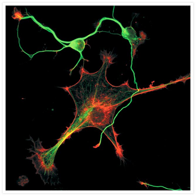
Cytoskeleton of a mixed population of granule neurons and glial cells
Confocal micrograph of the cytoskeleton of a mixed population of granule neurons and glial cells. The F-actin was stained with red-fluorescent Texas Red®-X phalloidin (Cat. No. T7471). The microtubules were detected with a mouse monoclonal anti–ß-tubulin primary antibody and subsequently visualized with the green-fluorescent Alexa Fluor® 488 Goat Anti–Mouse IgG antibody (Cat. No. A11001). The image was contributed by Jonathan Zmuda, Immunomatrix, Inc.

Related Products
Related Images
A prometaphase muntjac skin fibroblast stained with Alexa Fluor® 350 phalloidin, an anti–a-tubulin antibody and an anti–cdc6 peptide antibody. Go ›

Bovine pulmonary artery endothelial cells (BPAEC). MitoTracker® Red CMXRos, SYTOX® Green nucleic acid stain, biotin-XX goat anti–mouse IgG antibody and Cascade Blue® NeutrAvidin biotin-binding protein. Go ›

Endothelial cells. Go ›

Mouse Anti-Alpha Tubulin Monoclonal Antibody (Cat. No. A11126) Go ›

CD335 (NKp46) Antibody (63335182) in RE Go ›

CD223 (LAG-3) Antibody (56223942) in TM Go ›

Developing Drosophila embryo Go ›

Immunohistochemistry using GFAP Monoclonal Antibody, Mouse, Alexa Fluor® 594 Conjugate Go ›

Muntjac fibroblast labeled with probes for actin and the nucleus. Go ›

Multicolor fluorescence analysis of muntjac fibroblasts. Go ›
