Search Thermo Fisher Scientific
Pseudocolored micrograph of a single living smooth muscle cell labeled with a reduced form of rhod-2 AM.
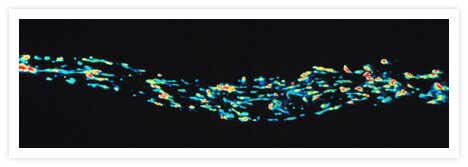
Pseudocolored micrograph of a single living smooth muscle cell labeled with a reduced form of rhod-2 AM (Cat. no. R1244, R1245MP), dihydrorhod-2. Images were acquired at focal planes spaced at 0.25 µm intervals and then processed using a constrained iterative deconvolution algorithm. This image shows that the rhod-2 fluorescence primarily arises from the mitochondria. The image was contributed by Fredric S. Fay, Program in Molecular Medicine, University of Massachusetts Medical Center.
Related Products
Related Images
Live cell imaging with CellLight™ reagents. Go ›

Live cells transduced with Organelle Lights™ or Cellular Lights™ reagents. Go ›

CD335 (NKp46) Antibody (63335182) in RE Go ›

CD223 (LAG-3) Antibody (56223942) in TM Go ›

REF-52 fibroblasts. Cyclic AMP Fluorosensor (FlCRhR) and fura-2 AM Go ›

Pseudocolored images of changes in intracellular free Ca2+ in AtT-20/D16v-F2 cells, monitored at 9 sec intervals with fluo-4, AM (F14201, F14202, F14217, F23917). Go ›
