Search Thermo Fisher Scientific
Invitrogen
CD56 (NCAM) Monoclonal Antibody (TULY56), PerCP-eFluor™ 710, eBioscience™
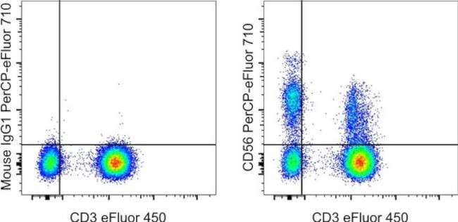
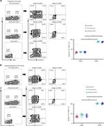
FIGURE: 1 / 11
CD56 (NCAM) Antibody (46-0566-42) in Flow


Product Details
46-0566-42
Species Reactivity
Published species
Host/Isotype
Recommended Isotype Control
Class
Type
Clone
Conjugate
Excitation/Emission Max
Form
Concentration
Purification
Storage buffer
Contains
Storage conditions
Shipping conditions
RRID
Product Specific Information
This TULY56 monoclonal antibody reacts with human CD56, also known as Neural Cell Adhesion Molecule (NCAM). CD56 is a highly glycosylated transmembrane molecule expressed by neurons and plays a role in the homotypic adhesion of neural cells. In the hematopoietic system, CD56 is expressed on NK cells and a subset of T cells referred to as NKT cells. Staining with TULY56 does not block binding of CMSSB, suggesting that the two antibodies recognize different epitopes. Additionally, TULY56 performs better after fixation and permeabilization than CMSSB.
The TULY56 monoclonal antibody crossreacts with Rhesus macaque.
This TULY56 antibody has been pre-titrated and tested by flow cytometric analysis of normal human peripheral blood cells. This can be used at 5 µL (0.06 µg) per test. A test is defined as the amount (µg) of antibody that will stain a cell sample in a final volume of 100 µL. Cell number should be determined empirically but can range from 10^5 to 10^8 cells/test.
PerCP-eFluor® 710 emits at 710 nm and is excited with the blue laser (488 nm); it can be used in place of PerCP-Cyanine5.5. We recommend using a 710/50 bandpass filter, however, the 695/40 bandpass filter is an acceptable alternative. Please make sure that your instrument is capable of detecting this fluorochrome.
Light sensitivity: This tandem dye is sensitive to photo-induced oxidation. Please protect this vial and stained samples from light.
Fixation: Samples can be stored in IC Fixation Buffer (Product # 00-8222) (100 µL of cell sample + 100 µL of IC Fixation Buffer) or 1-step Fix/Lyse Solution (Product # 00-5333) for up to 3 days in the dark at 4°C with minimal impact on brightness and FRET efficiency/compensation. Some generalizations regarding fluorophore performance after fixation can be made, but clone specific performance should be determined empirically.
Filtration: 0.2 µm post-manufacturing filtered.
Target Information
CD56 (NCAM, neural cell adhesion molecule) is a transmembrane glycoprotein of the immunoglobulin family that serves as an adhesive molecule and is ubiquitously expressed in the nervous system in isoforms ranging from 120-180 kDa. CD56 is found on T cells and NK cells, and is involved in cell migration, axonal growth, pathfinding and synaptic plasticity. Polysialic modification results in reduction of CD56-mediated cell adhesion. Through its extracellular region, CD56 mediates homophilic and heterophilic interactions by binding extracellular matrix components such as laminin and integrins. CD56 is expressed on most neuroectodermal derived cell lines, tissues and neoplasms such as retinoblastoma, medulloblastoma, astrocytomas and neuroblastoma. Further, CD56 is a widely used neuroendocrine marker with a high sensitivity for neuroendocrine tumors and ovarian granulosa cell tumors. Diseases associated with CD56 dysfunction include rabies and blastic plasmacytoid dendritic cell neoplasms.
For Research Use Only. Not for use in diagnostic procedures. Not for resale without express authorization.
How to use the Panel Builder
Watch the video to learn how to use the Invitrogen Flow Cytometry Panel Builder to build your next flow cytometry panel in 5 easy steps.
Bioinformatics
Protein Aliases: antigen recognized by monoclonal antibody 5.1H11; CD-56; CD56; E NCAM; N CAM1; N-CAM-1; neural cell adhesion molecule; Neural cell adhesion molecule 1; neural cell adhesion molecule, NCAM; sCD56; sNCAM; soluble CD56; soluble NCAM
Gene Aliases: CD56; MSK39; NCAM; NCAM1
UniProt ID: (Human) P13591
Entrez Gene ID: (Human) 4684, (Rhesus monkey) 693789

Performance Guarantee
If an Invitrogen™ antibody doesn't perform as described on our website or datasheet,we'll replace the product at no cost to you, or provide you with a credit for a future purchase.*
Learn more
We're here to help
Get expert recommendations for common problems or connect directly with an on staff expert for technical assistance related to applications, equipment and general product use.
Contact tech support
