Search Thermo Fisher Scientific
- Order Status
- Quick Order
-
Don't have an account ? Create Account
Search Thermo Fisher Scientific
In recent years, single-particle analysis (at both cryogenic and ambient temperatures) has emerged as a mainstream structural biology technique for the 3D characterization of macromolecules, proteins, and protein complexes at near or at atomic resolution. In fact, the 2017 Nobel Prize in Chemistry recognized the breakthrough developments in cryo-EM that make single particle analysis possible. Decades of dedicated work have refined the hardware, automation, and software needed to bring modern, accessible, and reliable single particle cryo-EM to the structural biology community and beyond.
Single particle cryo-EM or single particle analysis is an increasingly popular cryo-EM technique that allows you to investigate biomolecules at near- and atomic resolutions, unraveling dynamic biological processes and the structure of biomolecular complexes/assemblies. In single particle cryo-EM, purified proteins or protein complexes are suspended in amorphous (vitreous) ice through rapid plunge freezing, which preserve the samples’ native structures. Transmission electron microscopy (TEM) is then used to collect numerous 2D snapshots of the samples. As the proteins are oriented randomly within the ice, these images show the sample at various angles, and can be recombined into a high-resolution 3D reconstruction of the sample.
One of the primary challenges with investigating dynamic biological processes is the inherent complexity of biological machinery. Traditional structural biology techniques have struggled with large and/or dynamic protein systems, requiring indirect observation or the study of fragments. Luckily, single particle cryo-EM has emerged as a well-suited approach for the direct determination of native function and dynamics in complex biological systems. Single particle analysis can validate your biochemistry work by showing the molecular details that underlie the interactions between proteins, small molecules, and post-translational modifications in large and dynamic protein systems at near-native conditions. These molecular details can reveal the mechanisms by which complex biological systems contribute to human health and disease.
Single particle cryo-EM is particularly suited for the study of membrane proteins, protein complexes, and macromolecular machines such as viruses, ribosomes, and proteasomes. For example, the human GABAA (gamma-aminobutyric acid type A) receptor is a small membrane protein and ligand-gated chloride-ion channel that mediates inhibitory neurotransmission. GABAA receptors are important therapeutic targets as they impact a variety of important signaling pathways. Due to GABAA's conformational flexibility, traditional methods have been unable to reveal its molecular mechanism of action. Single particle analysis, however, revealed the molecular details of this important receptor, and how they underpin its allosteric modulation.
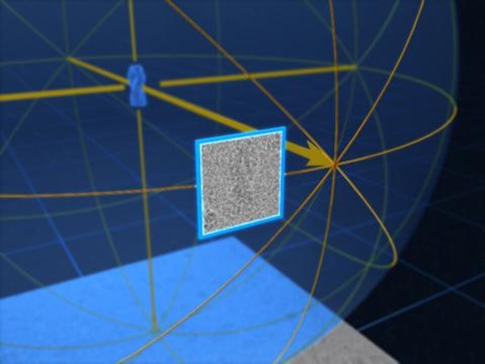
 Histamine in the binding pocket of human membrane protein, GABA-A receptor (resolved at 1.7 Å with the Krios Cryo-TEM). Data courtesy A. Sente and R. Aricescu, MRC-LMB Cambridge.
“We’ve been able to understand how these very few molecules are folded up and how they work, we can see where they change and where they can’t, and we can see exactly the shape that no one has ever seen before.”
Dr. Erica Saphire, La Jolla Institute of Immunology discussing the power of cryo-EM in studying viruses.
“The biggest advantage I see with cryo-EM…is that you need very little protein, so you are able to make reasonable amount in mammalian cells and without the need of crystals.”
Dr. Vinothkumar K.R, NCBS-TIFR, India
“Thanks to cryo-EM and thanks to single particle analysis we can directly extract those [amyloid] filaments from the heart of a patient who died from amyloidosis and we could get high resolution information about the structure and the overall architecture of the filament.”
Dr. Paolo Swuec, University of Milan, Italy, on discussing how he is using cryo-EM to better understand the pathology of amyloidosis


For Research Use Only. Not for use in diagnostic procedures.