Search Thermo Fisher Scientific
- Order Status
- Quick Order
-
Don't have an account ? Create Account
Search Thermo Fisher Scientific

Visit our Raman Spectroscopy Academy to learn the fundamentals of Raman spectroscopy and how you can apply this technology to your research, analyses, and quality assurance and quality control activities. Also, browse the resources at the bottom of this page to see how you can put Thermo Scientific Raman spectrometers and microscopes to work for your specific application.


Learn more about Raman microscopy and Raman spectroscopy, and the different applications the technology offers for analysis. You’ll find numerous different application notes from food and beverage to pharmaceuticals, as well as white paper, eBooks, and webinars.

Raman spectroscopy is a versatile vibrational technique and excels at identifying both organic and inorganic compounds in solids and liquids. Raman is non-destructive and requires little or no sample preparation.
We present an ongoing series of complimentary half-hour to one-hour webinars that reveal how the Thermo Scientific Raman instrumentation can improve your work.
Raman spectroscopy is essential to competitive academic research in many applied scientific disciplines, including materials science, life science research, and chemical and biological engineering. Advances in Raman microscopy and imaging have made the technique accessible to a wide variety of researchers, regardless of expertise or field of study.
During this webinar we will discuss:
Learn how Raman microscopy and imaging can help you answer your research questions.
Raman spectroscopy is rapidly gathering momentum as a valuable laboratory tool - an interesting development for a technique discovered in 1929. Join us to find out why this growth is happening - what made it possible - and to learn how to navigate the process of configuring your Raman system and then obtaining meaningful results. We will conclude by looking at some of the fields where Raman has made big inroads, like carbon materials (diamond, nanotubes, and graphene) and silicon.
During this webinar we will discuss:
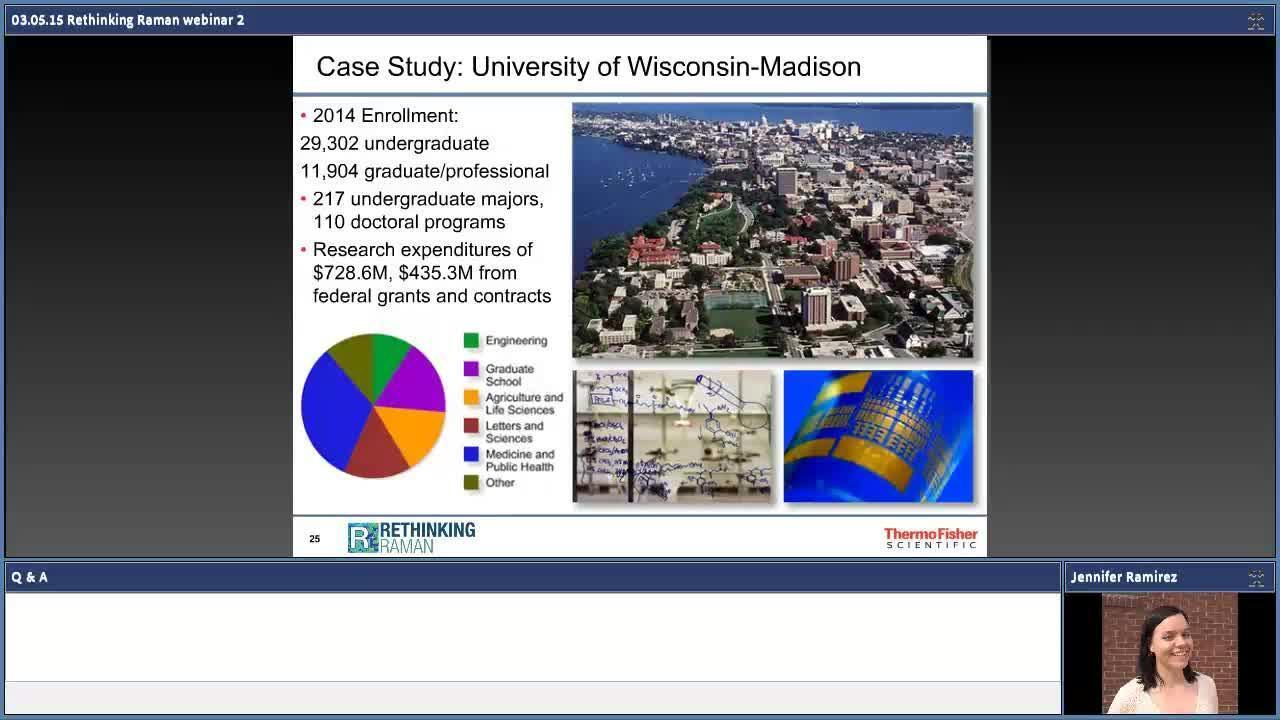
Rethinking Raman: Raman microscopy and imaging for shared academic labs
Raman spectroscopy is essential to competitive academic research in many applied scientific disciplines, including materials science, life science research, and chemical and biological engineering. Advances in Raman microscopy and imaging have made the technique accessible to a wide variety of researchers, regardless of expertise or field of study.
During this webinar we will discuss:
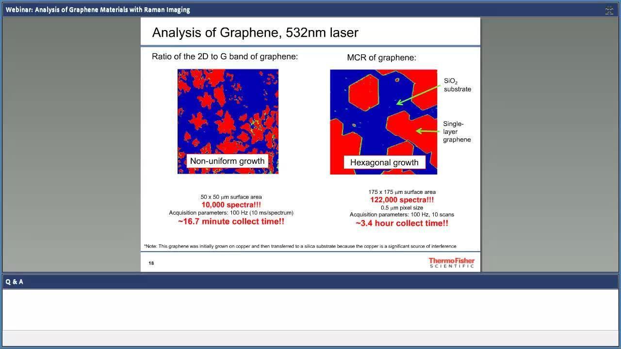
Graphene and based composites continue to be desirable materials due to mechanical, electrical and chemical properties. Potentially revolutionizing whole industries, from lighter-stronger advanced composites, flexible electronics, faster-smaller micro electronic devices, to more robust and efficient solar cell technologies, challenges associated with scaling up processes remain to be solved.
This webinar introduces how Raman imaging aids overcoming challenges associated with development of graphene and carbon nanotube based technologies. We will cover:
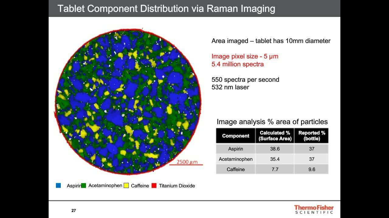
Pharmaceutical formations are typically complex mixtures that need to be carefully verified and understood. Raman spectroscopy is a proven method for identifying and verifying the presence of a variety of different components and providing detailed information on molecular structure and chemical environments. Raman imaging adds a spatial dimension to the analysis and extends the power of Raman spectroscopy across the sample.
Some of the applications of Raman imaging relevant to pharmaceutical evaluations include:
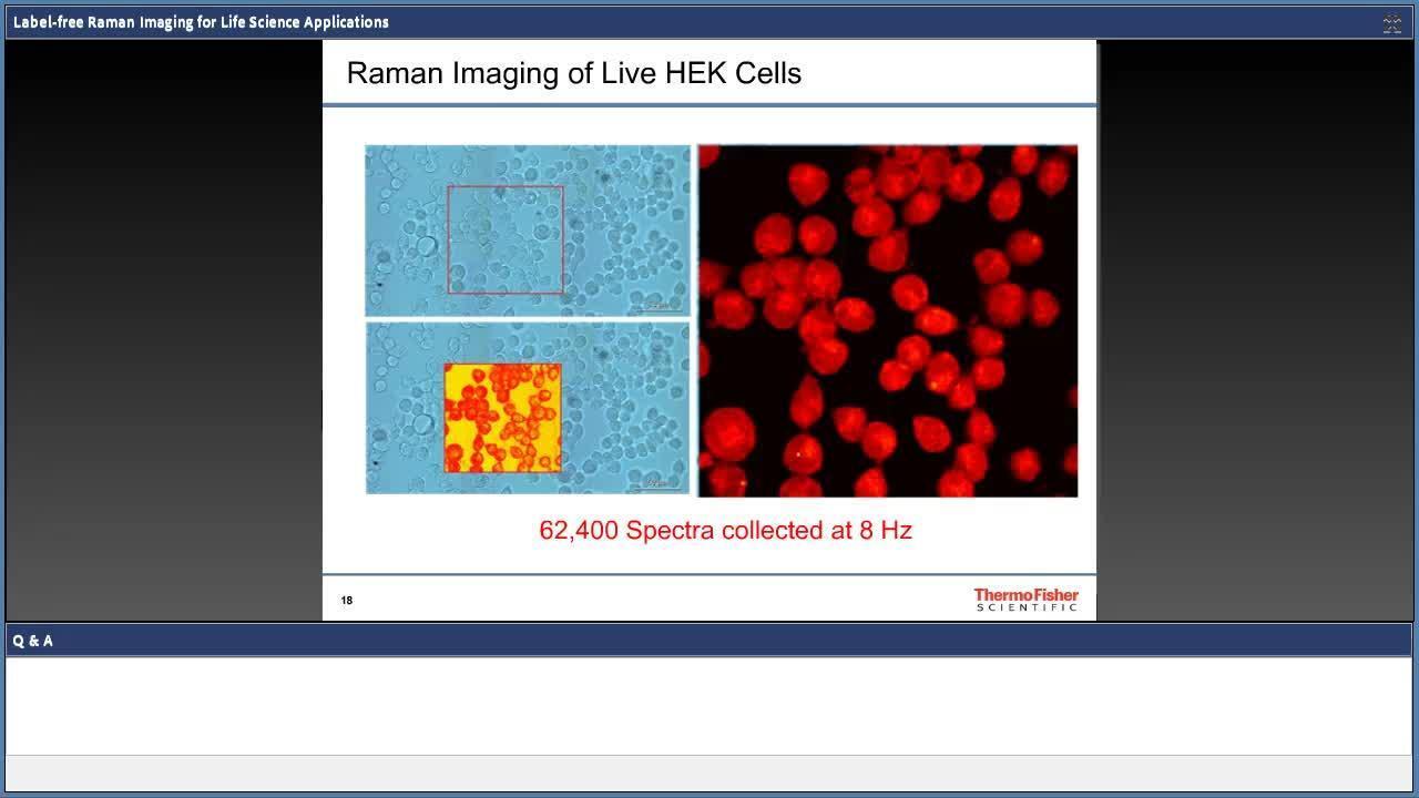
Raman imaging for life science research offers unique analytical capabilities for cell biology, enabling label-free characterization of biological systems with sub-micron spatial resolution. The ability to visualize living samples spatially and temporally to non-invasively understand molecular composition and dynamics has made Raman imaging a promising tool for cellular analysis. The DXRxi Raman imaging microscope allows the user to study living specimens through chemical imaging of components in their native environment. We will illustrate how this is an effective technique for live human cells, bacteria cells, and model organisms.
Among the topics to be discussed in this webinar:
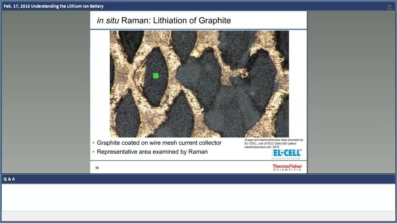
In our energy savvy society, we are seeing an increasing demand for renewable energy and new trends in energy storage. Although lithium-ion batteries offer the highest energy density among present commercial rechargeable batteries, the technology is still in need of vast improvements. Researchers seek solutions to understanding a diverse family of materials and chemical effects in lithium-ion batteries. Breaking through the ceiling of technology restraints is critical, and part of understanding the lithium-ion battery in full includes the chemistry inside of the cell.
This webinar illustrates how the lithium-ion battery works, and in effect, what types of problems are investigated within the laboratory. It will be the first part in a series of solution-focused webinars showing how we can help resolve the lithium-ion batteries' complicated mysteries.
What you will learn:
The rising demand of energy and the technological transformation of our energy systems require new and better battery materials. The success of the electric vehicle and the introduction of alternative energy sources are highly dependent on new batteries with higher energy density and longer life-time. Lithium-ion batteries are the actual state of technology, with a lot of potential for improvement and research beyond the lithium-ion technology, such as magnesium and sodium. The key for development is in new materials and the improvement of existing materials for better anodes, cathodes, separators, and electrolytes. However, before using these new materials in commercial batteries, it is necessary to characterize and test them in a laboratory environment.
This webinar introduces the basics of characterizing battery materials and the special equipment required to run these tests. We will specifically cover:
Basics of testing battery materials:
Exemplary case studies:
The relatively recent discovery of new allotropes of carbon, namely, carbon nanotubes and graphene, has opened up exciting new opportunities in the area of engineered materials. The properties that these materials can possess will be highly dependent upon the how these materials have been produced and/or modified.
Raman imaging: A tool for realizing graphene and graphene composite materials
Graphene and based composites continue to be desirable materials due to mechanical, electrical and chemical properties. Potentially revolutionizing whole industries, from lighter- stronger advanced composites, flexible electronics, faster-smaller micro electronic devices, to more robust and efficient solar cell technologies, challenges associated with scaling up processes remain to be solved.
This webinar introduces how Raman imaging aids overcoming challenges associated with development of graphene and carbon nanotube-based technologies. We will cover:
This presentation will focus upon what information is present in the Raman spectrum of carbon nanotubes and graphene and how Raman spectroscopy is a critical tool for everyone involved in the development and use of these materials.
This presentation will focus on characterizing graphene, carbon nanotubes, and diamond-like carbon films using Raman spectroscopy.
Learn how to:
Raman spectroscopy can characterize nanomaterials at different stages in their development, including guiding new synthetic routes for higher quality materials to assessing the effectiveness of desired chemical modifications and evaluating finished devices. This webinar will present a survey of applications where Raman gives important insight into these quickly evolving materials.
Raman spectroscopy provides important information such as layer thickness, uniformity, quality and functionalization, key parameters that ultimately determines the properties these materials will possess. This webinar will present an overview of recent work where Raman spectroscopy is playing vital role in realizing the development and full potential of these materials, spanning the range of applications from advanced composites, energy storage, transparent electrodes, and sensor technologies.
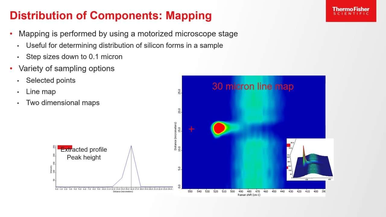
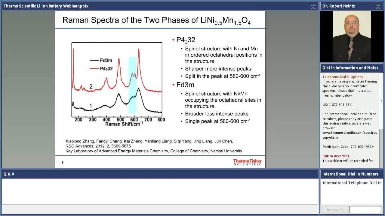
In our mobile society, we heavily rely on portable energy sources leading to driving improvements in battery technology. Although lithium-ion batteries offer the highest energy density among present commercial rechargeable batteries, the technology is still evolving and improving. Raman spectroscopy is a very versatile analytical tool that can be used to analyze the diverse materials that are used in lithium-ion batteries.
This presentation will illustrate how the structural and chemical information obtained from Raman spectroscopy can be applied to the analysis of components of lithium-ion batteries including cathodes, anodes, and electrolytes.
Good materials characterization is required across all steps in the creation of new graphene devices—from guiding the initial graphene synthesis, transfer to the desired substrate, and understanding chemical modification and analysis of the finished device. Our webinar presentation shows how a multi-technique approach using both Raman spectroscopy and XPS can address the challenges posed at these steps.
Using both techniques together allows analysts to completely characterize carbon nanomaterials. Our webinar demonstrates the utility of these techniques, illustrated by examples from graphene samples created by mechanical exfoliation, chemical reduction and CVD methods.
Areas of Interest:
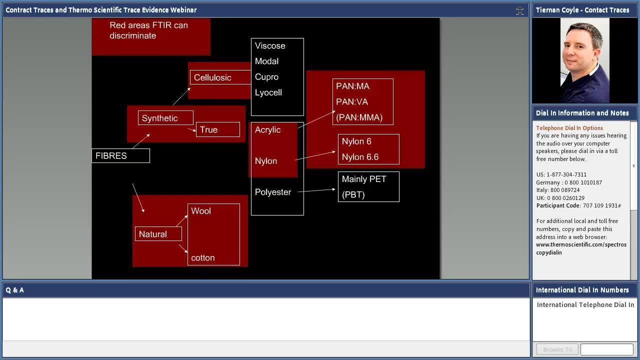
Trace evidence analysis: Raman and FTIR working together
Trace evidence examiners are required to obtain as much information as possible from microscopic amounts of material whilst respecting the need to maintain the integrity of the samples for any future forensic requirements. This webinar will demonstrate how spectroscopy plays a role in assisting the courts to answer some very difficult questions, with specific examples including additional levels of discrimination of fibers, glass; DNA –friendly methods of condom lubricant analysis and the potential for further discrimination of gun-shot residue.
This webinar demonstrates how new tools like enhanced low-pressure diamond cells and micro-ATR produce excellent results on samples important in Forensic investigations.
Topics will include:
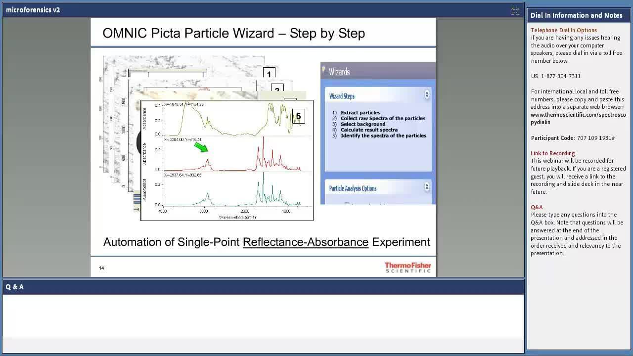
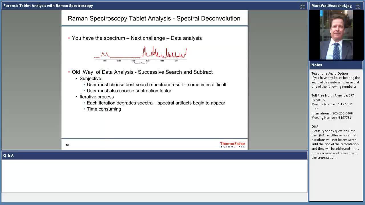
Raman spectroscopy can be used for the analysis of prescription, counterfeit, and designer drug tablets in just minutes with little to no sample preparation. As part of the presentation, we will make our debut presentation of the new Law Enforcement and Security (LEnS) spectral library which contains over 8300 unique spectra. Also featured will be the use of our multicomponent spectral search software, OMNIC Specta, for simplifying the analysis of complex tablet data.
Supplemental detection and classification methods of glaucoma disease states using Raman spectroscopy
This webinar discusses the potential of using Raman spectroscopy as a supplemental detection tool of glaucomatous changes in retinal tissues in vitro. Raman spectroscopic imaging was conducted on normal retinal tissues and retinal tissues with a variety of symptoms related to glaucoma. This webinar will address the use of Raman spectroscopy in clinical research, screening for the detection and characterization of early-stage glaucoma disease.
This webinar discusses:
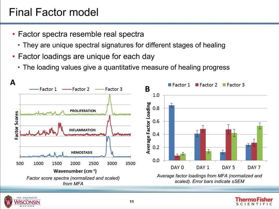
Biological tissues pose an analytical challenge because they are compositionally complex and the information, they provide can be difficult to objectively uncover. Raman spectroscopy offers an objective technique for characterization, however information from Raman spectra can be hard to reliably extract. We will explore how Raman microscopy can be utilized as a simple, non-invasive method to biochemically characterize healing wounds, particularly when coupled with multivariate spectral analysis to accurately identify different phases of the healing of wounds with time in clinical research.
Considering wounds as a model biological surface, the webinar will address:
Raman imaging for life science research offers unique analytical capabilities for cell biology, enabling label-free characterization of biological systems with sub-micron spatial resolution. The ability to visualize living samples spatially and temporally to non-invasively understand molecular composition and dynamics has made Raman imaging a promising tool for cellular analysis. The DXRxi Raman imaging microscope allows the user to study living specimens through chemical imaging of components in their native environment. We will illustrate how this is an effective technique for live human cells, bacteria cells, and model organisms.
Among the topics to be discussed in this webinar:
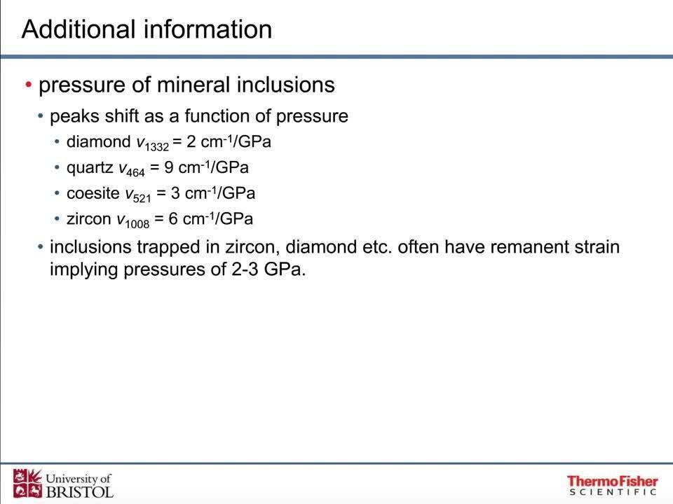
Raman and infrared microscopy of minerals and fluid inclusions
Infrared and Raman spectroscopy are convenient and information-rich analytical techniques for characterizing a wide variety of samples important in earth science. Combined with microscopy, vibrational spectra can identify specific minerals, characterize contents of fluid inclusions, and produce chemical images of complex mixtures. This presentation discusses how infrared and Raman complement other micro-techniques such as optical microscopy and SEM-EDS. We will show how Raman microscopy can deliver key data not possible by any other technique.
Raman spectroscopy can provide information that is complementary to other techniques, or it might be the only feasible method of analysis in a conventional laboratory. Learn how Raman spectroscopy can be used routinely to non-destructively study geological samples.
Topics covered will include:
Visualizing, characterizing, and analyzing pharmaceutical constituents with Raman imaging
Pharmaceutical formations are typically complex mixtures that need to be carefully verified and understood. Raman spectroscopy is a proven method for identifying and verifying the presence of a variety of different components and providing detailed information on molecular structure and chemical environments. Raman imaging adds a spatial dimension to the analysis and extends the power of Raman spectroscopy across the sample.
Some of the applications of Raman imaging relevant to pharmaceutical evaluations include:
The combination of FTIR and Raman spectroscopy with mapping is well-suited for identifying and sizing API domains. Both techniques provide excellent chemical and spatial information about the distribution and concentration of the API in a tablet.
During this webinar, you’ll learn how to:
Biopharmaceutical manufacturers are increasing their testing regimes and increasing their efforts to raise current production levels. However, due to bottlenecks in current testing procedures, it’s not always easy to shorten testing times. This webinar addresses the role of the Raman spectroscopy in the rapid release of raw materials through identity testing and real-time release testing of final products such as vaccines, hormones, and monoclonal antibodies.
Key learning objectives
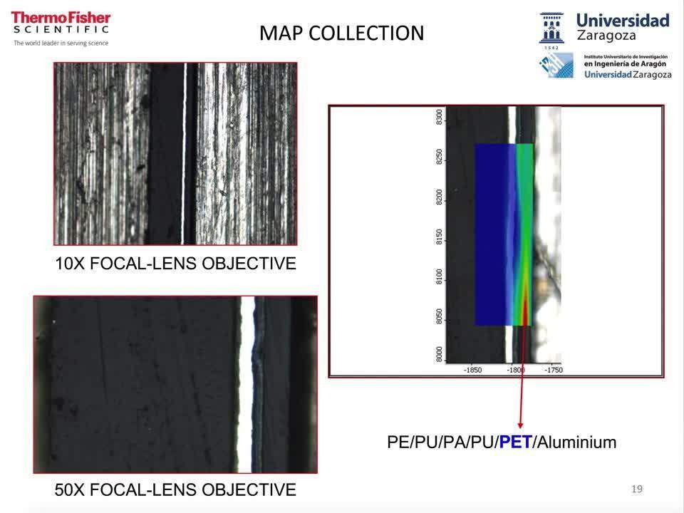
Your next packaging analysis tool – Raman microscopy
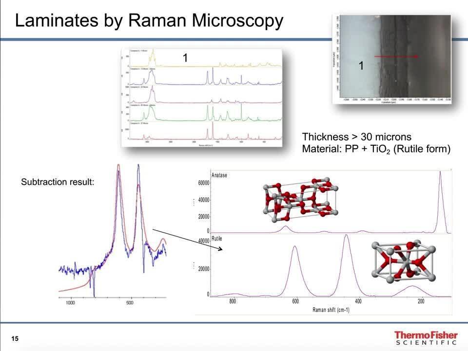
Raman microscopy is a valuable tool for packaging analysis, with special emphasis on food-contact materials. In minutes, you can characterize multilayer material properties such as structure and thickness and can even identify layers with minimal sample handling. In this webinar, we discuss novel promising applications of Raman microscopy, such as:
Raman microscopy is a powerful technique that can be utilized for the analysis of multilayer polymer films, thus enabling the control of composition and quality. Conventional Raman microscopy, which has spatial resolution as small as a micron, can be employed to analyze cross sections of multilayer polymer films. Confocal Raman microscopy can be used in situations where a reduction in sample preparation is desired, as it can generate depth profiles of the multilayer films, with no requirement for cross sectioning.
For Research Use Only. Not for use in diagnostic procedures.