Search Thermo Fisher Scientific
- Order Status
- Quick Order
-
Don't have an account ? Create Account
Search Thermo Fisher Scientific
 Generation of reactive oxygen species (ROS) is inevitable for aerobic organisms and, in healthy cells, occurs at a controlled rate. Under conditions of oxidative stress, ROS production is dramatically increased, resulting in subsequent alteration of membrane lipids, proteins, and nucleic acids. Oxidative damage of these biomolecules is associated with aging as well as a variety of pathological events, including atherosclerosis, carcinogenesis, ischemia reperfusion injury, and neurodegenerative disorders.
Visualize mitochondrial superoxide in your live cells with specificity and ease-of-use, MitoSOX Green offers:
We provide a variety of Invitrogen fluorescent tools to track different parameters in oxidative stress:
These tools use platforms such as fluorescence microscopy, flow cytometry, or microplate analysis.
Select products for oxidative stress detection
Detection of oxidative stress with Invitrogen™ CellROX™ Green Reagent in U2OS cells.
Oxidative stress results from an imbalance in the production of reactive oxygen species (ROS) and the ability of the cell to scavenge them. ROS react with nucleic acids, proteins and lipids causing cell and tissue damage and can be measured using selective or general indicators.
| Readout | CellROX Dyes are non-fluorescent in a reduced state and exhibit bright, stable fluorescence upon oxidation by reactive oxygen species (ROS). They are membrane permeant and load readily into live cells | Non-fluorescent dye becomes green when oxidized | ||
| Common filter set | Cy5 | RFP | FITC | FITC |
| Reporter | CellROX Deep Red Reagent | CellROX Orange Reagent | CellROX Green Reagent | H2-DCFDA |
| Ex/Em (nm) | 640/665 | 545/565 | 485/520 | 495/527 |
| Live cell–compatible | Yes | Yes | Yes | Yes |
| Labeling in complete medium | Yes | Yes | Yes | No |
| Formaldehyde-fixable | Yes | No | Yes | No |
| Detergent-resistant | No | No | Yes | No |
| Photostability |  | |  |  |
| Signal-to-noise ratio | | | | |
| Bibliography | Citations | Citations | ||
| Imaging | Yes | Yes | Yes | Yes |
| HCS | Yes | Yes | Yes | Yes |
| Microplate | Yes | Yes | Yes | Yes |
| Flow cytometry | C10491 (kit) | C10493 (kit) | C10492 (kit) | Yes |
| Format | 5 x 50 uL | 5 x 50 uL | 5 x 50 uL | 20 x 50 ug |
| Cat. No. | C10422 | C10443 | C10444 | C6827 |
Lipid peroxidation is the oxidative degradation of lipids. Reactive oxygen species are the major initiators of lipid peroxidation and membrane bound polyunsaturated fatty acids like arachidonic acid and linoleic acid are their major targets. The byproducts of lipid peroxidation cause direct damage to cell membranes. They also form protein adducts resulting in cell and tissue damage. Lipid peroxidation is implicated in many human diseases including diabetes and cardiovascular disease.
Image-iT Lipid Peroxidation Kit,
for live cell analysis | |||
|---|---|---|---|
| Readout | Ratiometric indicator of lipid peroxidation shifts from red to green fluorescence with oxidation | Single wavelength indicator detects lipid-peroxidation derived protein modifications in fixed cells | |
| Common filter set(s) | FITC and Texas Red | FITC | |
| Fluorophore | BODIPY 581/591 C11 | Alexa Fluor 488 | |
| Ex/Em (nm) | 495/519 | 581/591 | 495/519 |
| Live cell–compatible | Yes | No | |
| Detects fixed cells | No | Yes | |
| Whole cell imaging | Yes | Yes | |
| Live cell–compatible | Yes | No | |
| Photostability |  | | |
| Signal-to-noise ratio | | | |
| Bibliography | Citations | Citations | |
| Formaldehyde-fixable | No | Detects fixed cells | |
| Antibody-multiplexable/ Detergent-resistant | No | Yes | |
| Imaging | Yes | Yes | |
| HCS | Yes | Yes | |
| Microplates | Yes | Yes | |
| Flow cytometry | Yes | Yes | |
| Format | 500 assay kit | 500 assay kit | |
| Cat. No. | C10445 | C10446 | |
Superoxide, peroxyl radical, hydrogen peroxide, hydroxyl radical and peroxynitrite are some examples of ROS that react with nucleic acids, proteins and lipids and result in cell and tissue damage. Certain ROS have been implicated in various human diseases including cancer, cardiovascular disease, neurodegenerative disease and aging.
| Dihydroethidium | Premo Cellular Hydrogen Peroxide (H2O2) Sensor | ||||
|---|---|---|---|---|---|
| Specificity | Superoxide (•O2–) | Superoxide (•O2–) | Superoxide (•O2–) | Nitric Oxide (NO) | Hydrogen peroxide (H2O2) |
| Readout | Targeted to mitochondria
| Targeted to mitochondria | Blue fluorescent dye becomes red when oxidized | Fluorescence increases with accumulation of NO | Detects H2O2 levels with ratio of excitation at 400 and 488 nm and single emission at 515 nm |
| Common filter set | Custom Filter set | FITC | RFP | FITC | None |
| Fluorophore | MitoSOX Red | MitoSOX Green | Dihydroethidium (DHE) | DAF-FM | roGFP |
| Ex/Em (nm) | 396/610 | 488/510 nm | 518/606 | 495/515 | 400 and 488/515 |
| Live cells | Yes | Yes | Yes | Yes | Yes |
| Fixable | No | No | Yes | No | No |
| Labeling in complete medium | No | No | No | Yes | |
| Photostability | | | | ||
| Signal-to-noise ratio | | | | ||
| Bibliography | Citations | Citations | Citations | Citations | |
| Imaging | Yes | Yes | Yes | Yes | Yes |
| HCS | Yes | Yes | Yes | Yes | Yes |
| Microplate | Yes | Not Tested | Yes | Yes | |
| Flow Cytometry | Yes | Not Tested | Yes | Yes | |
| In vivo | Yes | No | |||
| Format | 10 x 50 ug | 5 x 9 µg | 10 x 1 mg | 10 x 50 ug | 5 mL |
| Cat. No. | M36008 | M36006 | D11347 | D23844 | P36243 |
*It should be noted that two excitation peaks may be observed for MitoSOX Red, as it is excited at both 510 nm and 396 nm. While 510 nm will excite superoxide oxidation product, it can also excite non-specific products so we recommend using a 396 nm excitation for more selective detection of mito superoxide when using MitoSOX Red.
Reduced glutathione also known as GSH is a major thiol bound to proteins. Protein thiols including GSH play an important role in determining the redox status of cells. Therefore, detection of reduced GSH levels is a useful indication of redox potential and a cell's ability to prevent oxidative stress.
| Readout | Intracellular probe for GSH used in subcellular detection and localization as well as monitoring of sub populations | Reacts with several low molecular weight thiols to generate fluorescent conjugates | Detects redox potential with ratio of excitation wavelength and single emission | ||
| Common filter set | DAPI longpass | DAPI | None | ||
| Ex/Em (nm) | 404/525 | 394/490 | 400 and 488/515 | ||
| Fluorophore | ThiolTracker Violet | Monochlorobimane (mBCI) | Monobromobimane (mBBr) | roGFP | |
| Live cell– compatible | Yes | Yes | Yes | ||
| Formaldehyde-fixable | Yes | No | No | ||
| Antibody-multiplexable | Yes | No | No | ||
| Whole cell imaging | Yes | Yes | Yes | ||
| Photostability | |||||
| Signal-to-noise ratio | |||||
| Bibliography | Citations | Citations | Citations | ||
| Imaging | Yes | Yes | Yes | ||
| HCS | Yes | Yes | Yes | ||
| Microplates | Yes | Yes | Yes | Yes | |
| Flow cytometry | Yes | Yes | Yes | Yes | |
| Format | 180 assay kit | 500 assay kit | 25 mg | 25 mg | 5 mL |
| Cat. No. | T10095 | T10096 | M1381MP | M1378 | P36424 |
Oxidative stress results from an imbalance in the production of reactive oxygen species (ROS) and the ability of the cell to scavenge them. ROS react with nucleic acids, proteins and lipids causing cell and tissue damage and can be measured using selective or general indicators.
| Readout | CellROX Dyes are non-fluorescent in a reduced state and exhibit bright, stable fluorescence upon oxidation by reactive oxygen species (ROS). They are membrane permeant and load readily into live cells | Non-fluorescent dye becomes green when oxidized | ||
| Common filter set | Cy5 | RFP | FITC | FITC |
| Reporter | CellROX Deep Red Reagent | CellROX Orange Reagent | CellROX Green Reagent | H2-DCFDA |
| Ex/Em (nm) | 640/665 | 545/565 | 485/520 | 495/527 |
| Live cell–compatible | Yes | Yes | Yes | Yes |
| Labeling in complete medium | Yes | Yes | Yes | No |
| Formaldehyde-fixable | Yes | No | Yes | No |
| Detergent-resistant | No | No | Yes | No |
| Photostability | | | | |
| Signal-to-noise ratio | | | | |
| Bibliography | Citations | Citations | ||
| Imaging | Yes | Yes | Yes | Yes |
| HCS | Yes | Yes | Yes | Yes |
| Microplate | Yes | Yes | Yes | Yes |
| Flow cytometry | C10491 (kit) | C10493 (kit) | C10492 (kit) | Yes |
| Format | 5 x 50 uL | 5 x 50 uL | 5 x 50 uL | 20 x 50 ug |
| Cat. No. | C10422 | C10443 | C10444 | C6827 |
Lipid peroxidation is the oxidative degradation of lipids. Reactive oxygen species are the major initiators of lipid peroxidation and membrane bound polyunsaturated fatty acids like arachidonic acid and linoleic acid are their major targets. The byproducts of lipid peroxidation cause direct damage to cell membranes. They also form protein adducts resulting in cell and tissue damage. Lipid peroxidation is implicated in many human diseases including diabetes and cardiovascular disease.
Image-iT Lipid Peroxidation Kit,
for live cell analysis | |||
|---|---|---|---|
| Readout | Ratiometric indicator of lipid peroxidation shifts from red to green fluorescence with oxidation | Single wavelength indicator detects lipid-peroxidation derived protein modifications in fixed cells | |
| Common filter set(s) | FITC and Texas Red | FITC | |
| Fluorophore | BODIPY 581/591 C11 | Alexa Fluor 488 | |
| Ex/Em (nm) | 495/519 | 581/591 | 495/519 |
| Live cell–compatible | Yes | No | |
| Detects fixed cells | No | Yes | |
| Whole cell imaging | Yes | Yes | |
| Live cell–compatible | Yes | No | |
| Photostability | | | |
| Signal-to-noise ratio | | | |
| Bibliography | Citations | Citations | |
| Formaldehyde-fixable | No | Detects fixed cells | |
| Antibody-multiplexable/ Detergent-resistant | No | Yes | |
| Imaging | Yes | Yes | |
| HCS | Yes | Yes | |
| Microplates | Yes | Yes | |
| Flow cytometry | Yes | Yes | |
| Format | 500 assay kit | 500 assay kit | |
| Cat. No. | C10445 | C10446 | |
Superoxide, peroxyl radical, hydrogen peroxide, hydroxyl radical and peroxynitrite are some examples of ROS that react with nucleic acids, proteins and lipids and result in cell and tissue damage. Certain ROS have been implicated in various human diseases including cancer, cardiovascular disease, neurodegenerative disease and aging.
| Dihydroethidium | Premo Cellular Hydrogen Peroxide (H2O2) Sensor | ||||
|---|---|---|---|---|---|
| Specificity | Superoxide (•O2–) | Superoxide (•O2–) | Superoxide (•O2–) | Nitric Oxide (NO) | Hydrogen peroxide (H2O2) |
| Readout | Targeted to mitochondria
| Targeted to mitochondria | Blue fluorescent dye becomes red when oxidized | Fluorescence increases with accumulation of NO | Detects H2O2 levels with ratio of excitation at 400 and 488 nm and single emission at 515 nm |
| Common filter set | Custom Filter set | FITC | RFP | FITC | None |
| Fluorophore | MitoSOX Red | MitoSOX Green | Dihydroethidium (DHE) | DAF-FM | roGFP |
| Ex/Em (nm) | 396/610 | 488/510 nm | 518/606 | 495/515 | 400 and 488/515 |
| Live cells | Yes | Yes | Yes | Yes | Yes |
| Fixable | No | No | Yes | No | No |
| Labeling in complete medium | No | No | No | Yes | |
| Photostability | | | | ||
| Signal-to-noise ratio | | | | ||
| Bibliography | Citations | Citations | Citations | Citations | |
| Imaging | Yes | Yes | Yes | Yes | Yes |
| HCS | Yes | Yes | Yes | Yes | Yes |
| Microplate | Yes | Not Tested | Yes | Yes | |
| Flow Cytometry | Yes | Not Tested | Yes | Yes | |
| In vivo | Yes | No | |||
| Format | 10 x 50 ug | 5 x 9 µg | 10 x 1 mg | 10 x 50 ug | 5 mL |
| Cat. No. | M36008 | M36006 | D11347 | D23844 | P36243 |
*It should be noted that two excitation peaks may be observed for MitoSOX Red, as it is excited at both 510 nm and 396 nm. While 510 nm will excite superoxide oxidation product, it can also excite non-specific products so we recommend using a 396 nm excitation for more selective detection of mito superoxide when using MitoSOX Red.
Reduced glutathione also known as GSH is a major thiol bound to proteins. Protein thiols including GSH play an important role in determining the redox status of cells. Therefore, detection of reduced GSH levels is a useful indication of redox potential and a cell's ability to prevent oxidative stress.
| Readout | Intracellular probe for GSH used in subcellular detection and localization as well as monitoring of sub populations | Reacts with several low molecular weight thiols to generate fluorescent conjugates | Detects redox potential with ratio of excitation wavelength and single emission | ||
| Common filter set | DAPI longpass | DAPI | None | ||
| Ex/Em (nm) | 404/525 | 394/490 | 400 and 488/515 | ||
| Fluorophore | ThiolTracker Violet | Monochlorobimane (mBCI) | Monobromobimane (mBBr) | roGFP | |
| Live cell– compatible | Yes | Yes | Yes | ||
| Formaldehyde-fixable | Yes | No | No | ||
| Antibody-multiplexable | Yes | No | No | ||
| Whole cell imaging | Yes | Yes | Yes | ||
| Photostability | |||||
| Signal-to-noise ratio | |||||
| Bibliography | Citations | Citations | Citations | ||
| Imaging | Yes | Yes | Yes | ||
| HCS | Yes | Yes | Yes | ||
| Microplates | Yes | Yes | Yes | Yes | |
| Flow cytometry | Yes | Yes | Yes | Yes | |
| Format | 180 assay kit | 500 assay kit | 25 mg | 25 mg | 5 mL |
| Cat. No. | T10095 | T10096 | M1381MP | M1378 | P36424 |
Invitrogen CellROX Reagents are fluorogenic probes for measuring generalized oxidative stress in cells using conventional fluorescence microscopy, high content screening, microplate fluorometry, or flow cytometry. The dyes are nonfluorescent in a reduced state and fluoresce bright green, orange, or deep red upon oxidation. Some are formalin fixable for easy multiplexing with other fluorescent reagents including antibodies. Assays are simple and reliable with a sensitive and robust readout, and the reagent can be applied to cells in complete growth media. We provide Invitrogen CellROX dyes and conventional probes for oxidative stress measurement.
|  General oxidative stress detection in U2OS cells with CellROX Green Reagent. |
Lipid peroxidation is the oxidative degradation of lipids. ROS are the major initiators of lipid peroxidation, and membrane-bound polyunsaturated fatty acids like arachidonic acid and linoleic acid are their major targets. The byproducts of lipid peroxidation cause direct damage to cell membranes. They also form protein adducts, resulting in cell and tissue damage. Lipid peroxidation is implicated in many human diseases including diabetes and cardiovascular disease. We provide Invitrogen tools for live and fixed cells, with a choice of ratiometric or single-wavelength readout. |  Detection of lipid peroxidation with the Invitrogen Image-iT Lipid Peroxidation Kit. |
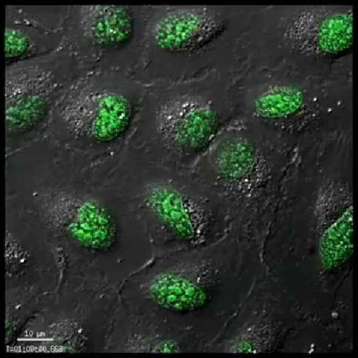
Superoxide, peroxyl radical, hydrogen peroxide, hydroxyl radical, and peroxynitrite are examples of ROS that react with nucleic acids, proteins, and lipids and result in cell and tissue damage. Certain ROS have been implicated in various human diseases including cancer, cardiovascular disease, neurodegenerative disease, and aging. We provide selective Invitrogen reagents for a variety of reactive species targeted to the cytosol or mitochondria and detected using imaging, flow cytometry, or microplate analysis. |  Detection of superoxide in live cells using Invitrogen MitoSOX Red Superoxide Indicator. |
Genetically encoded sensors provide an alternative to organic dyes for measuring redox potential or oxidative stress in live cells. roGFP is a fluorescent protein chimera that changes its excitation maximum from 488 nm to 400 nm based on a highly efficient redox relay. Using ratiometric detection of the emission at 515 nm, this shift can be used to measure H2O2 (Invitrogen Premo Cellular Hydrogen Peroxide H2O2 Sensor) or redox potential (Invitrogen Premo Cellular Redox Sensor, Grx-1-roGFP) in live cells in a reversible and reproducible assay. The fluorescent proteins can be multiplexed with organic dyes or other expressed proteins to provide additional biological context for the assay. |  Detecting changes in cellular redox potential in U2OS cells using Premo Cellular Redox Sensor. |
Reduced glutathione, also known as GSH, is a major thiol bound to proteins. Protein thiols including GSH play an important role in determining the redox status of cells. Therefore, detection of GSH levels is a useful indication of redox potential and a cell’s ability to prevent oxidative stress. We provide Invitrogen intracellular probes for the sensitive detection and localization of thiols. |  Detection of GSH in U2OS cells with Invitrogen ThiolTracker Violet dye. |
For Research Use Only. Not for use in diagnostic procedures.
