Search Thermo Fisher Scientific
Volumescope 2 Scanning Electron Microscope for serial block face imaging
The Thermo Scientific Volumescope 2 Scanning Electron Microscope (SEM) is our state-of-the-art serial block-face imaging system. Keep control of experiments with easy to use technology and protect a wide range of possible samples with tried and tested solutions for every acquisition step.
Block face imaging
Unraveling the complex 3D architecture of cells and tissues in their natural context is crucial for structure-function correlation in biological systems and soft materials. Serial block-face SEM (SBF-SEM) combines in situ sectioning and imaging of plastic-embedded tissue blocks within the SEM vacuum chamber for automated, 3D reconstruction of large tissue volumes. Axial resolution was once limited by the minimum section thickness, but truly isotropic resolution is now possible with the addition of multi-energy deconvolution.
3D SEM
Following in situ sectioning of the block-face using a diamond knife, the freshly exposed tissue is imaged several times using increasing accelerating voltages. These images are subsequently used in a deconvolution algorithm to derive several optical subsurface layers, forming a 3D subset. By repeating this cycle, the Volumescope 2 SEM offers isotropic datasets with 10 nm Z-resolution.


Polymer microstructures
High-resolution imaging in 3D is of great importance when trying to understand the microstructure and properties of polymer materials. The Volumescope 2 SEM combines in situ sectioning and imaging of polymers within the SEM vacuum chamber, in a fully automated fashion, for reconstruction of large volumes with truly isotropic 3D resolution. Visualizing a large volume at such resolutions is critical for revealing how regions with different microstructures affect the overall properties of the material. For example, the Volumescope 2 SEM can recreate a large representative volume of a filtration membrane, providing accurate transport properties that lead to enhanced performance predictions for new filter designs.

Easy to use technology
Keep control of experiments with easy to use technology. Reuse jobs and system settings and select multiple regions of interest during job acquisition.
Visualize and navigate during acquisition
Visualize and navigate during acquisition with Thermo Scientific Amira Live Tracker Software to optimize/control your outcome/Automate large 3D volume acquisitions as well as reconstructions.
Protect valuable samples
Protect valuable samples with tested solutions at every acquisition step: debris trap and swipe features ensure sample quality; low vacuum detector enables imaging of highly charged samples.
Easy and fast mounting microtome exchange
Easy and fast mounting microtome exchange for normal SEM operation or automated array tomography with the addition of Thermo Scientific Maps Software.
| Electron optics |
|
| Source lifetime | 24 months |
| Automation |
|
| Stage | Double stage scanning deflection |
| Electron beam |
|
| Chamber |
|
| Detectors |
|
| Vacuum system |
|
| Sample holders |
|
See datasheet for full list of specifications including software, accessories and a summary of installation requirements.

Pesquisa de patologia
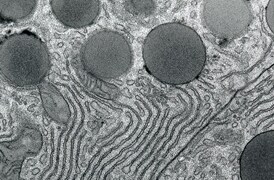
A microscopia eletrônica de transmissão (TEM) é usada quando a natureza da doença não pode ser estabelecida por métodos alternativos. Com a geração de imagens nanobiológicas, a TEM fornece informações precisas e confiáveis para determinadas patologias.

Pesquisa de biologia vegetal
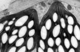
A pesquisa fundamental da biologia vegetal é possível devido à microscopia crioeletrônica. Ela fornece informações sobre proteínas (com análise de partículas únicas), sobre seu contexto celular (com tomografia), até a estrutura geral da planta (análise de volume grande).

Pesquisa de materiais fundamentais
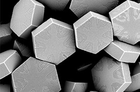
Novos materiais são investigados em escalas cada vez menores para o máximo controle de suas propriedades físicas e químicas. A microscopia eletrônica fornece aos pesquisadores percepções importantes sobre uma ampla variedade de características materiais em escala micro a nano.

Controle de qualidade
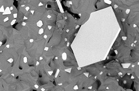
O controle de qualidade e a garantia de qualidade são essenciais na indústria moderna. Oferecemos uma gama de ferramentas de microscopia eletrônica e espectroscopia para análises multidimensionais e multimodais de defeitos, permitindo que você tome decisões confiáveis e informadas para controle e melhoria de processos.

Análise de grande volume
Uma nova solução de imagens de face de bloco serial (SBFI) que combina microscopia eletrônica de varredura de deconvolução de várias energias (MED-SEM) com seccionamento in situ. As funções de automação e a facilidade de uso fornecem resolução isotrópica para amostras de grande volume.
Serviços de microscopia eletrônica
Para garantir o desempenho ideal do sistema, fornecemos acesso a uma rede de especialistas em serviços de campo, suporte técnico e peças de reposição certificadas.


