Search Thermo Fisher Scientific
Iliad (S)TEM features for advanced materials analysis
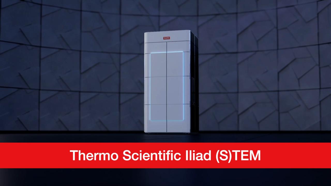
With advanced integration, the Thermo Scientific Iliad (S)TEM can help you answer challenging scientific questions and surpass previous limitations. Advanced spectroscopy capabilities along with dose optimization strategies make it ideal for complex investigations of even the most difficult modern materials.
Advanced optics integration for superior EELS performance
EELS optics have the challenge of simultaneously transferring a broad range of electron energies through the microscope and spectrometer, from specimen to detector, without introducing chromatic blur or chromatic distortions. This is a non-trivial task, especially when trying to maintain the transfer across different microscope settings and across different spectrometer settings.
Atomically resolved EELS elemental map of LaMnO3/LaFeO3 interface.
This can only be ensured by closely integrating the optics of the microscope and spectrometer to match the chromatic defocus in the microscope with the focus of the spectrum. Otherwise, the superior performance offered by the tapered prism and ten multipoles could only be fully enjoyed at one or a few specific experimental settings and would be lost when switching to other camera lengths, dispersions, or energy offsets. Our EELS Spectrometer and Energy Filter, combined with advanced optics and high stability, offers unique integration with the TEM optics to improve the EELS data collection experience.
Atomically resolved EELS elemental map of TbScO3 with Tb shown in green and Sc shown in red.
NanoPulser Electrostatic Beam Blanker
An electrostatic beam blanker is used in electron microscopy to optimize electron dose. It selectively blocks or blanks the beam, effectively turning it on and off over the specimen at a high frequency. TEMs typically use magnetic deflectors, which only have a response time of milliseconds and are not as reproducible as electrostatic deflectors. The Nanopulser — an electrostatic beam blanker — enables precise temporal control over the electron dose delivered to the sample and supports a wide range of applications including, for example, dose-optimized investigations.
Electron beam scanning over the specimen with (left) the conventional STEM settings (no blanking) and (right) pulsating beam while beam blanker is on providing 50% blanking of beam during scan and completely blanked flyback signal.
Velox Software
Cutting-edge Thermo Scientific Velox Software for transmission electron microscopy offers comprehensive experimental control. It facilitates access to scanning transmission electron microscope (STEM and TEM) optics and detectors, enhancing reproducibility, yield, and support for quantitative STEM and TEM material analysis.
Velox Software stands out with its integrated ergonomic user interface and ease of use, providing high quality in imaging and compositional mapping. The integrated SmartCam brings efficient setup of experiments. Additionally, it has an interactive detector layout interface for optimal experimental control and documentation on multidetector tools. Velox Software supports high-contrast atomic imaging of light and heavy elements and enables flexible STEM and TEM movie recording for dynamic studies.
Enhancing EELS and EDS spectroscopy with Velox Software
Velox Software is equipped with unique packages for electron energy loss spectroscopy and energy-dispersive X-ray spectroscopy on Thermo Scientific EDX detectors. This robust mapping engine integrates multiple techniques optimized for transmission electron microscopy, ensuring acquisition of best-in-class spectrum images with high yield. The software also provides live feedback during acquisition and fast post-processing of EELS and EDS data.
Overall, Velox Software is a powerful tool that offers excellent control, quality, and versatility in transmission electron microscopy, making it an essential part of the scientific research ecosystem.
High energy electron resolution sources
The Iliad (S)TEM offers advanced configuration options with high-energy-resolution X-FEG/Mono or ultra-high-energy-resolution X-FEG/UltiMono energy sources. Both sources automatically tune for optimal energy resolution necessary for EELS experiments. Operating from 30 – 300 kV, they accommodate diverse experiments, including STEM-EDS mapping, EELS, and ultra-high-resolution STEM.
Cold field emission gun
The Iliad (S)TEM offers the option of a cold field emission gun (X-CFEG) with high brightness and low energy spread. It operates from 30 – 300 kV, providing high-resolution STEM imaging with high probe currents. The X-CFEG allows flexible tuning of probe currents and energy resolution, enabling a wide range of experiments. Tip flashing is required once per working day, with no impact on probe aberrations or tip lifetime. The X-CFEG also supports standard TEM imaging experiments with large parallel probes.
STEM imaging performance
The Iliad (S)TEM, equipped with either the X-FEG/Mono or X-FEG/UltiMono energy source, achieves the highest commercially available STEM resolution specifications. With enhanced mechanical stability, fifth-order probe aberration correction, and a high-resolution (S-TWIN) wide-gap pole piece, it achieves resolutions of 50 pm at 300 kV, 96 pm at 60 kV, and 125 pm at 30 kV with 30 pA or 100 pA probe current (with X-CFEG).
Panther STEM detection system
STEM imaging on the Iliad (S)TEM is transformed with the Panther STEM detection system. Featuring two new solid-state detectors with 16 segments, this system offers advanced imaging capabilities and single-electron sensitivity. Optimized signal chain and low-dose imaging enable beam-sensitive material imaging while the scalable architecture allows for combining detector segments and synchronizing multiple signals for innovative STEM techniques.
4D STEM cameras
The Iliad (S)TEM can be configured with an electron microscope pixel array detector (EMPAD) or a Thermo Scientific Ceta Camera with speed enhancement for 4D STEM data collection.
The EMPAD provides high dynamic range, signal-to- noise ratio, and speed, making it optimal for 4D STEM applications. The Ceta Camera offers higher resolution diffraction patterns and is suitable for applications such as strain measurement and EDS analysis. For more information, please refer to the EMPAD datasheet.
Lorentz microscopy
The Iliad (S)TEM can be used to study pristine magnetic structures in a field-free mode. Configurable with virtually zero magnetic field across the sample volume, it can conduct experiments in both TEM and STEM modes. This enables high-resolution studies such as DPC or ptychography with high resolution. It can also perform in situ magnetization to observe the behavior of structures under real operating conditions.
For Research Use Only. Not for use in diagnostic procedures.
