Search Thermo Fisher Scientific

Remarkable image quality for a range of applications
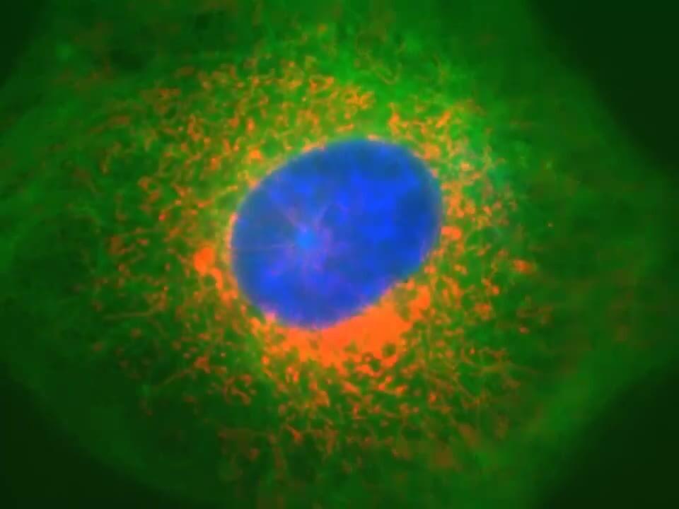
Explore our collection of publication-quality images, videos, and applications data generated with EVOS imaging systems.
EVOS image gallery
The central feature shared by all EVOS cell imaging systems is their remarkable image quality. In just a few clicks, capture clear, bright, publication-quality images that help you tell the story of your data. This collection of images demonstrates the versatility and range of applications that EVOS imaging systems can help visualize. Filter by the EVOS model or dye used. View the antibodies used in the immunostaining.
Instrument
Dye
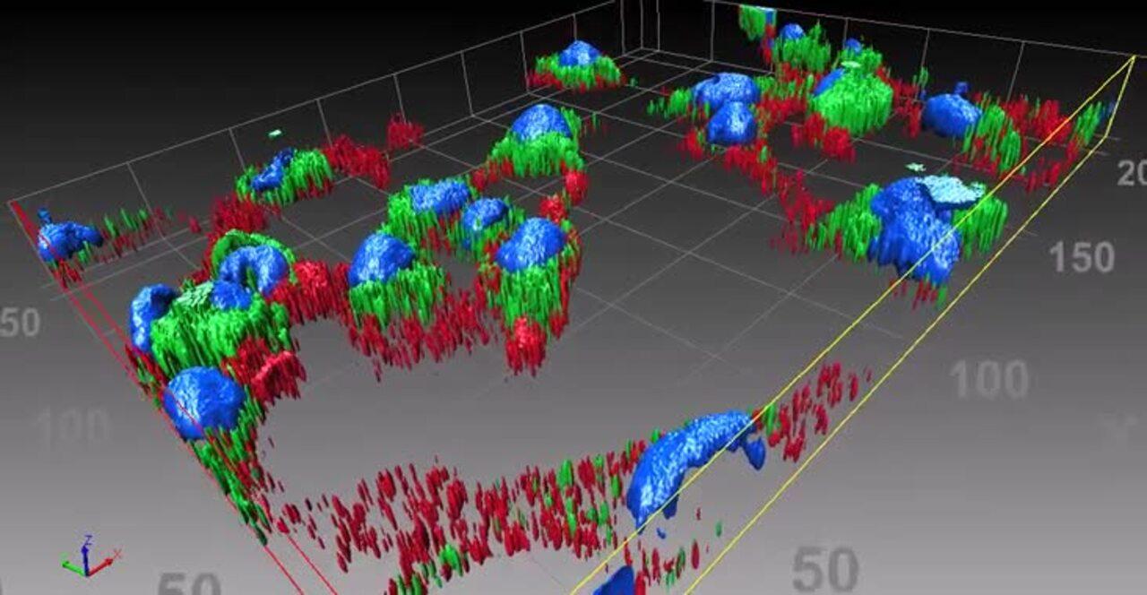
EVOS video gallery
With EVOS imaging systems, you can compile movies from individual images, such as time-lapse, Z-stack, tile-stitching, whole plate stitching, 3D visualization, analysis, and deconvolution. The gallery below demonstrates some of the ways that EVOS videos can help you tell the story of your data.


Applications data
EVOS imaging systems support a broad range of imaging applications—including cell culture, time-lapse imaging, and high-resolution capture from slides, dishes, flasks, and microplates. Below you will find featured application data which is available as application notes and/or scientific posters.


Reference articles
Designed to eliminate the complexities of microscopy without compromising performance, EVOS imaging systems make cell imaging accessible to almost every lab and budget. Use our Research Publications Database to learn how researchers like you are using EVOS imaging systems to publish their results.

For Research Use Only. Not for use in diagnostic procedures.