Search Thermo Fisher Scientific

Shared features of EVOS imaging systems
In today’s competitive scientific environment, generating publication-quality images is critical. Discover how EVOS imaging systems have been designed with enabling your success in mind, including outstanding imaging components, compact designs, and intuitive software.
High-resolution imaging
The ability to generate clear and detailed images is critical. High-resolution allows for outstanding visualization and analysis of cellular structures and processes. The central feature shared by all EVOS cell imaging systems is their remarkable image quality. In a few clicks, capture clear, bright, publication-quality images and videos that help you tell the story of your data.
Variable magnification options

EVOS cell imaging systems include a high-sensitivity monochrome camera, excellent for fluorescence imaging; a high-sensitivity color camera, exceptional for colorimetric imaging; or both monochrome and color cameras for maximum flexibility—depending on the model. EVOS objectives offer outstanding optical performance from visible light to near-infrared light.
Choose from more than 40 high-performance objective lenses ranging from 1.25x to 100x. Long working distance (LWD) objectives are optimized for vessels with a nominal wall thickness of 0.9–1.5 mm, such as slides, cell culture dishes and flasks, and microtiter plates. For applications using 1.5 coverslips (approximately 0.17 mm thick), coverslip-corrected (CC) objectives have a higher magnification-to-numerical aperture (NA) ratio and provide higher resolution than LWD objectives.
Exceptional fluorescence imaging and illumination

All EVOS imaging systems feature bright LED light sources that provide more than 50,000 hours of brilliant, yet adjustable illumination that, unlike mercury arc lamps, remains constant with minimal degradation over time. Cost-efficient and environmentally friendly, EVOS LED light cubes have been optimized to take cell imaging to the next level with precise control, minimal maintenance, and exceptional reliability.
A choice of compact, easily interchangeable LED light cubes can be matched to the spectra of specific dyes or fluorescent proteins in your application. Each cube incorporates excitation filter, dichroic beam-splitter, and emission filter. Filters are hard-coated to enable maximum transmission efficiency.
Compact, efficient design
EVOS microscopes are designed with scientists’ workspace and workflow needs in mind. Components and controls are conveniently integrated into a single, lightweight system that fits on a benchtop and can easily be moved to classrooms, teaching labs, and conference rooms. The size and portability of EVOS microscopes let you capture and view images when and where needed with no darkroom required.


Enhanced safety, minimal handling
The EVOS M3000 and EVOS M5000 imaging systems are designed for use in a biosafety cabinet or hood while wearing gowns, gloves, and goggles or face shields if necessary. There’s minimal risk of contamination when you passage, split, transfect, transduce, or otherwise manipulate your cells.
Further, with the advanced EVOS imaging systems, software controls most or all microscope operations. You can adjust focus, zoom, pan, and (with the EVOS M7000) even control stage movement remotely through the software without touching the instrument, helping minimize the possibility of infection or cross-contamination through physical contact. With the EVOS M7000, you can run automated routines such as multi-well plate scanning and successive scans of a designated area. The only required interaction with the instrument is to mount the slide, plate, or other vessel.

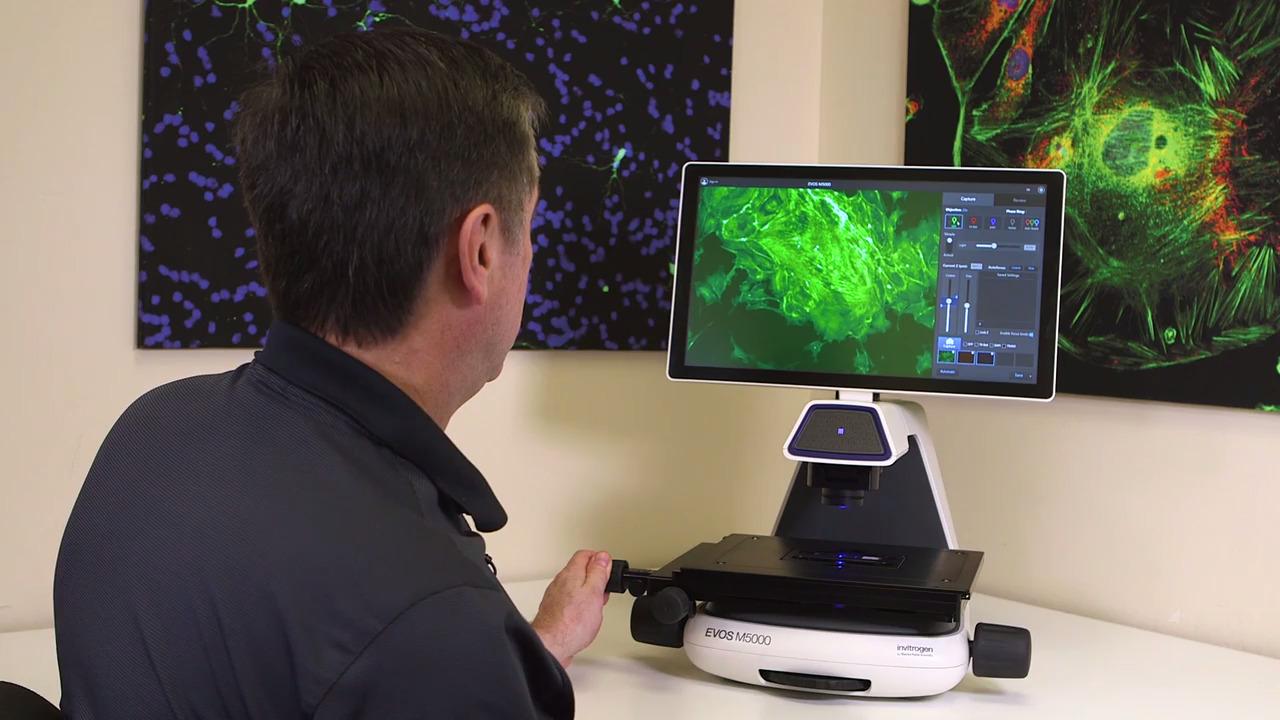
Powerful, intuitive software
All EVOS microscopes come with onboard software that allows you to view, capture, and process images quickly and easily. You can:
- select colors and adjust brightness while viewing an image
- adjust contrast, saturation, and color balance
- choose between high resolution and quick capture
- capture and save images
On the EVOS M7000 and EVOS M5000, the onboard software offers pinpoint operational control and powerful image processing tools such as cell counting, confluence, Z-stacking, image tiling and stitching, time-lapse movies, and RGB synthesis. These tools operate intuitively with minimal training, aided by onboard SmartStart orientation. Autofocus and other automated tools help save time and effort. The EVOS M7000 adds advanced automation features and complete programmatic control, allowing imaging through software alone, without ever touching the instrument.
Download the most recent EVOS M3000 software updates
Download the most recent EVOS M5000 software updates
Download the most recent EVOS M7000 software updates
Intuitive software user interface
All EVOS imaging systems share many characteristics that make them easy to learn and operate. Increasing the number of features doesn’t need to equal increased complexity. For every application, EVOS systems are designed with the user in mind, helping make even the most complicated workflows simple to perform. This is the EVOS system experience.


Celleste Image Analysis Software
For advanced image processing and analysis, the Celleste Image Analysis Software option offers powerful additional tools to segment, count, size, classify and analyze complex images, including machine learning algorithms for advanced image analysis and quantitation. Optional modules for deconvolution, 3D visualization, and 3D analysis allow you to customize the capabilities according to your needs.
Thermo Fisher cloud connection
Images from all EVOS instruments can be sorted and analyzed with the EVOS Image Analysis app in Connect, Thermo Fisher’s secure, cloud-based platform for data storage, scientific analysis apps, and peer collaboration tools. You can link your Thermo Fisher Connect account to an EVOS M5000 Imaging System in very few steps directly from the instrument, from a mobile device, or from your computer. After you have signed in to your Thermo Fisher Cloud account from the M5000 instrument, you can choose to upload captured images directly to the cloud application, in addition to local storage. Once uploaded, you and your colleagues can collaboratively access your gallery and retrieve, analyze, and edit your images from any web browser around the world. You can also run other Connect applications and monitor the status and configuration of your connected instruments.


In the Image Analysis app, you can zoom and pan images, adjust the brightness and contrast of each channel, and apply pseudocolors. You can also overlay a region and display its dimensions. When analyzing an image, once you identify your target cells vs background, the app can automatically count the cells in a given area and quantify their area, fluorescence intensity, and circularity.

For Research Use Only. Not for use in diagnostic procedures.