Search Thermo Fisher Scientific

Make way for more
Bring automated, fast, and high-performance imaging right to your bench with the EVOS M7000 Imaging System. This system has been designed with advanced capabilities to simplify demanding slide and cell-based imaging applications including live-cell analysis, image tiling, and Z-stacking, so you can focus on acquiring images and data rather than instrument operation.
Features
In addition to the common features of all EVOS microscopes, the EVOS M7000 Imaging System offers:
Full automation
Automated routines help streamline workflow and improve experimental reproducibility
Speed
Scan a 96-well plate in 3 fluorescence channels in less than 5 minutes
Two cameras, no compromises
Dedicated cameras for color and fluorescence produce high-resolution images and data
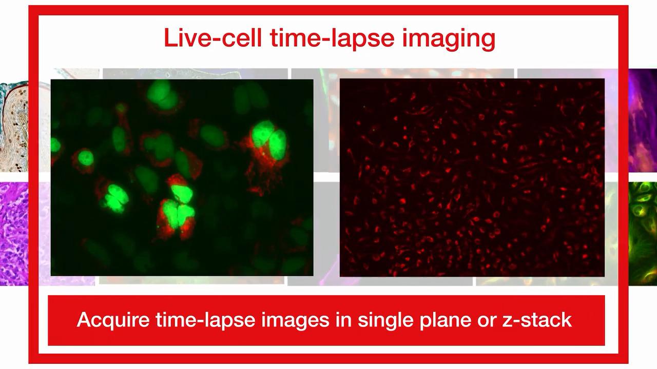
Time-lapse live-cell imaging
Optional onstage incubator enables precise control over temperature, humidity, and gas levels
Area view
Move rapidly and seamlessly between single-field mode and low- and high-magnification scan modes to easily define and capture the area of interest
Data analysis
Seamlessly transfer images to optional
Celleste Image Analysis Software for access to powerful tools for image segmentation, classification, and cell-based assays
Virtual 360º view
Check out the EVOS M7000 Imaging System virtually.
Explore 360º view

EVOS M7000 HCA Package
The EVOS M7000 Imaging System, High-Content Analysis Package (AMF7000HCA) combines the fully automated, inverted, multi-channel fluorescence and transmitted light imaging system with Celleste 6 Image Analysis Software, a full-featured image analysis suite. The Celleste 6 Image Analysis software facilitates the transition into low-throughput high-content analyses (HCA) while maintaining the versatility and power of the EVOS M7000 automated microscope.
Using plate-based multi-channel analysis (MCA) protocols leveraging machine-learning based algorithms together with its icon-based wizard-driven workflow, Celleste 6 software can help you efficiently segment and classify images for a wide range of assays or bioapplications, including neurite outgrowth, angiogenesis, cell viability, transfection efficiency, and many more. The software also offers a wide range of well-plate data displays including heat maps, image montages, and kinetic graphing options.
EVOS M7000 & Celleste 6 HCA Packages
Description |
Cat. No |
EVOS M7000 Imaging System, High Content Analysis Package
|
|
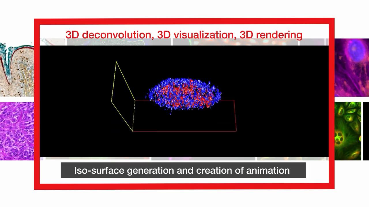
EVOS M7000 Imaging System, High Content Analysis Package with 3D
|
 Multi-class protocol for Celleste 6
Featured images and videos
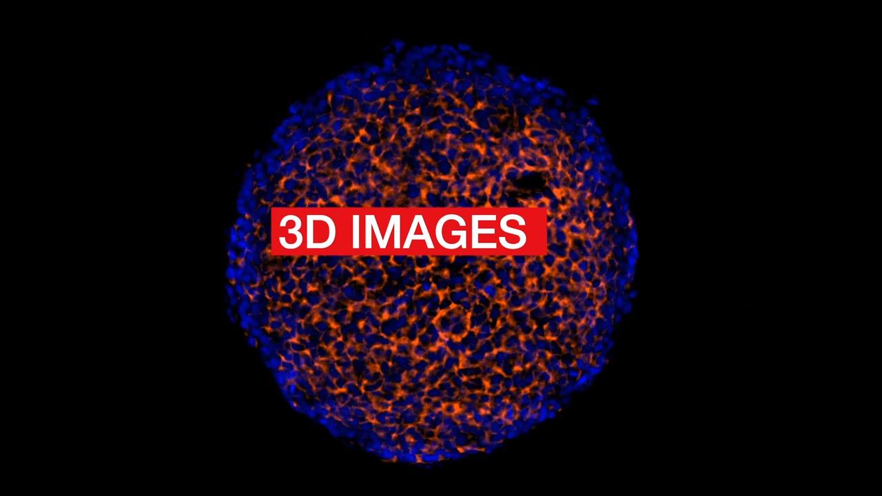


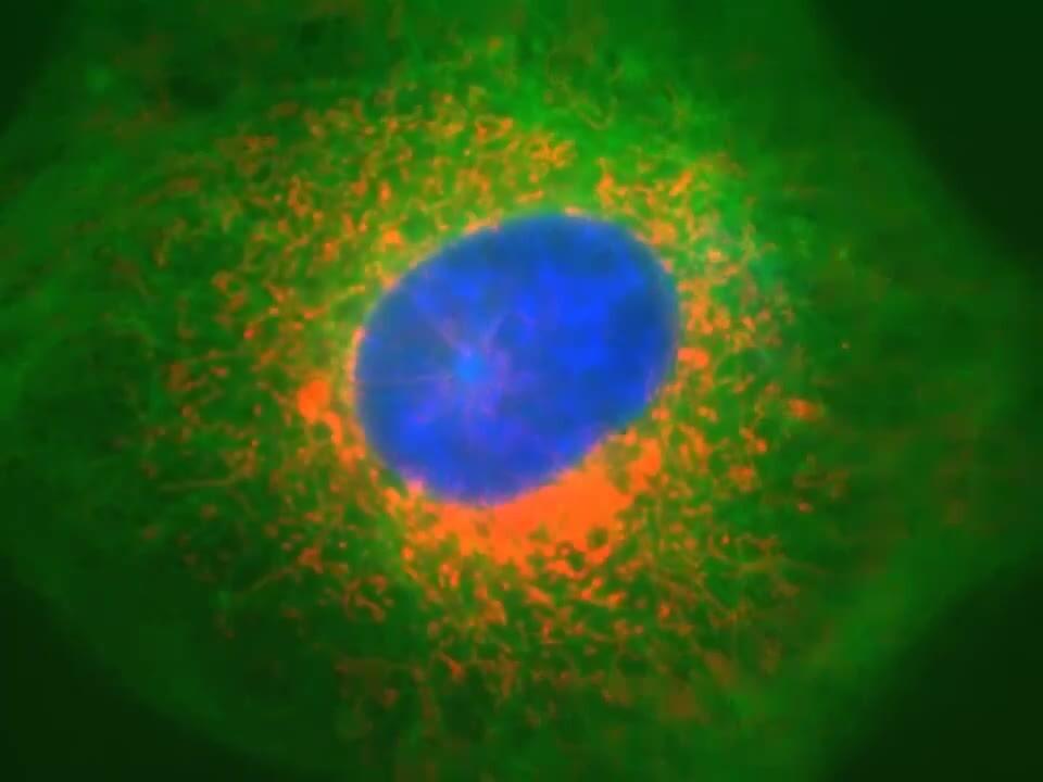

View more M7000 images and movies in our Image Gallery and Video Gallery.
Specifications
| Category | Attribute | Description |
| Optics | Description | Infinity-corrected optical system; RMS threaded objectives with 45 mm parfocal distance |
| Imaging mode | Fluorescence, brightfield, color brightfield, phase contrast | |
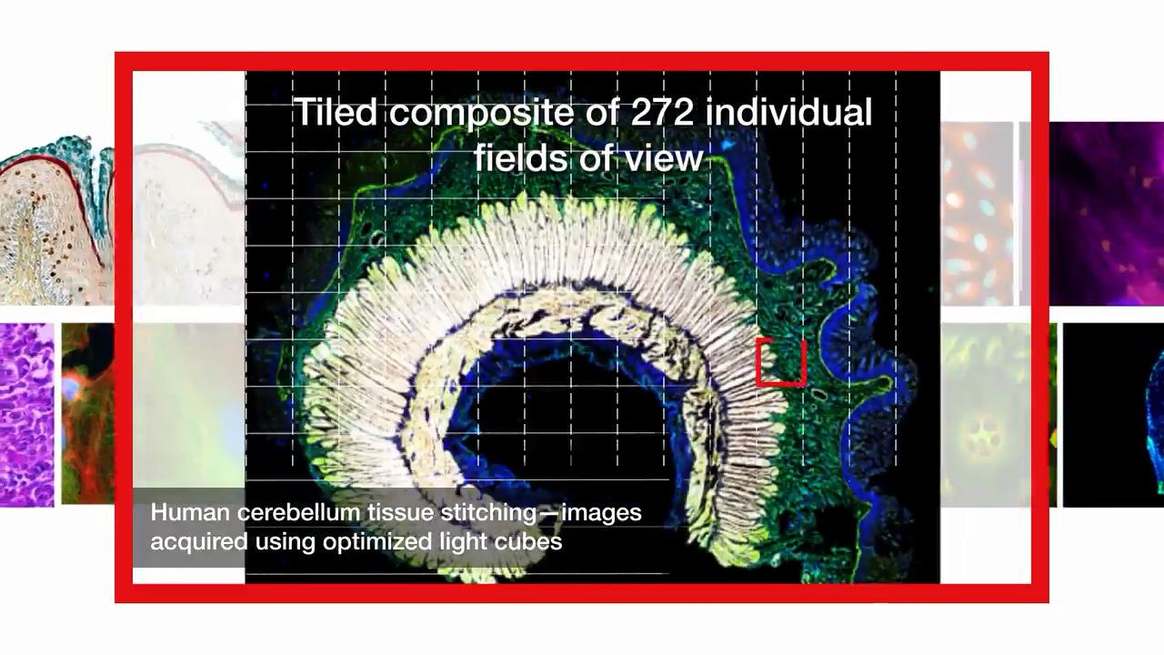
| Imaging methods | Single color, multicolor, area scan with montage or tile-stitch, time lapse, Z-stacking, movie capture | |
| Illumination | Adjustable-intensity LED light cubes with integrated hard-coated filter set and >50,000-hour life | |
| Light cube capacity | 5-position chamber for 4 fluorescence cubes + brightfield | |
| Light cubes (not included) | Broad selection of standard and specialty light cubes. Commonly used light cubes include (ex/em): • DAPI (357/447 nm) • GFP (470/525 nm) • RFP (531/593 nm) • Texas Red (585/624 nm) • Cy5 (628/692 nm) |
|
| Objective capacity | 5-position automated turret | |
| Objectives (not included) | Wide selection of high-quality long working distance (LWD) and coverslip-corrected objectives | |
| Condenser | 60-mm LWD condenser; 4-position turret with a clear aperture and 3 phase annuli | |
| Focus mechanism | Automated focus with sub-micron (0.150 µm) resolution (single-step accuracy) | |
| Monochrome camera | High-sensitivity 3.2 MP (2048 x 1536) CMOS sensor with 3.45 µm pixel resolution | |
| Color camera | High-sensitivity 3.2 MP (2048 x 1536) CMOS sensor with 3.45 µm pixel resolution | |
| Captured images | 16-bit RAW monochrome: TIFF, PNG (12-bit dynamic range) 8-bit color: TIFF, PNG, JPG Movies and time-lapse: AVI, WMV |
|
| Physical characteristics | Power supply | 24V AC adapter with country-specific power cords |
| Dimensions (W x H x D) | 18 x 14 x 13 inches 45.7 x 33.0 x 35.6 cm |
|
| Weight | 26 lbs (11.8 kg) | |
| X/Y scanning stage | Travel range 120 mm x 80 mm with sub-micron resolution Drop-in inserts to receive vessel holders and lock-down holders to fix sample in place during long scans |
|
| Stage mechanism | Motorized | |
| Automation | Plate scanning | Automated multiwell |
| System | Computer | External Dell XE4 computer with Intel Core i9 processor, 32 GB DDR4 RAM, 512 GB PCIe NVME SSD, and NVIDIA Quadro RTXA4000 with 8 GB discrete video graphics running Windows®10 |
| Storage | 32 GB DDR4 RAM 512 GB PCIe SSD |
|
| Output ports | Computer: 1 x USB 3.1 Gen 2 Type-C; 5 x USB 3.1 Gen 1 Type-A; 4 x USB 2.0 Type-A; 1 Serial; 2 Display Ports 1.2; 1 RJ-45; 2 PS/2; 1 UAJ; 1 Line-out | |
| Networking capability | Connection through Windows/SMB network via Ethernet cable connection |
For Research Use Only. Not for use in diagnostic procedures.