Search Thermo Fisher Scientific

Easily track and go back
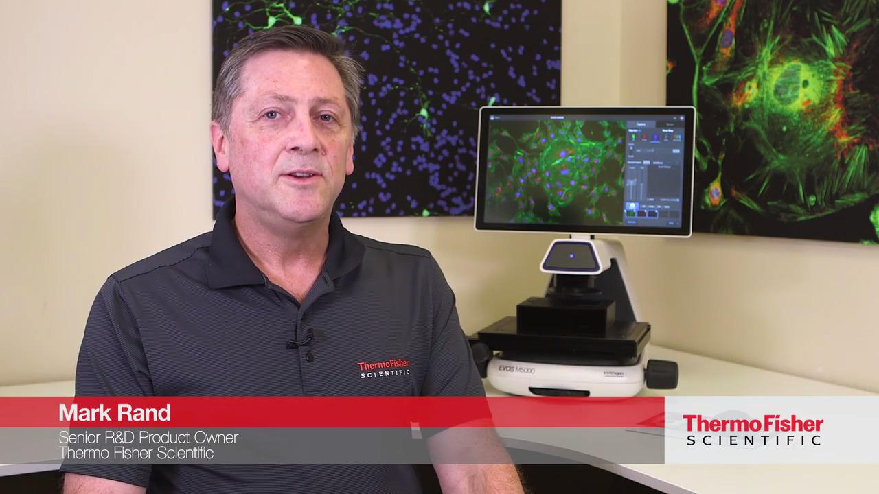
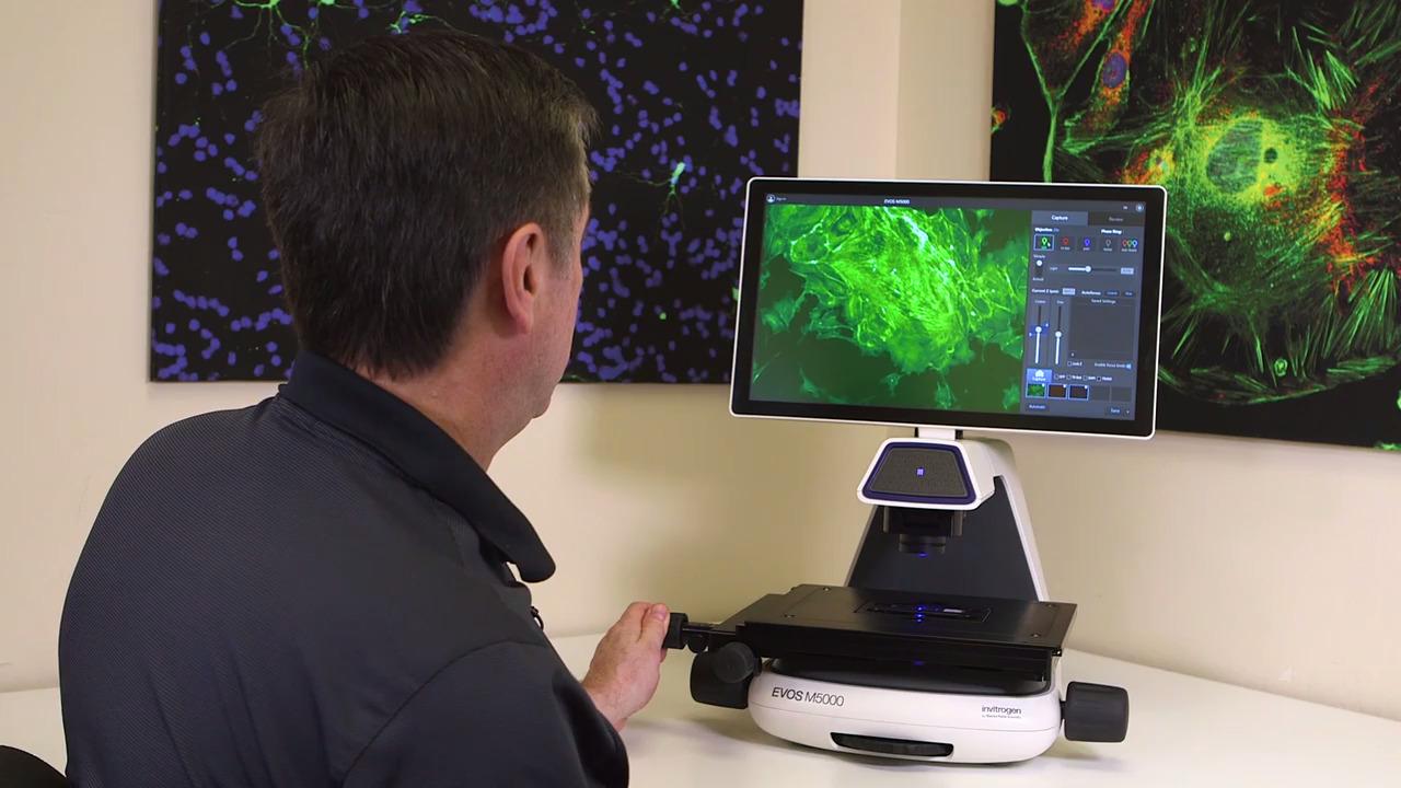
The fully integrated EVOS M5000 Imaging System combines precision optics, an articulated 18.5-inch high-resolution LCD monitor, and a highly sensitive camera. It delivers high-quality four-color fluorescence, transmitted light, phase-contrast, and color images with excellent flexibility across a broad range of applications and imaging requirements. Designed by biologists for biologists, this remarkably easy-to-use microscope enables seamless image acquisition and provides a convenient set of tools for analysis and annotation that can be used in live mode and with saved images.
The Stage View feature allows you to track your location as you manually move the stage and search for your cells. Areas of interest can be marked, saved, and returned to, at any time.
New stage-tracking intelligence
Watch this video to see how you can effortlessly locate, track, and record points of interest.
Features
In addition to the common features of all EVOS microscopes, the EVOS M5000 Imaging System offers:
Easily track and go back
Stage-tracking intelligence simplifies returning to precise stage locations
Optimal performance
Autofocus, Z-stack capability, time-lapse imaging, and multichannel capture with a single click
Onboard analytics
Easy access to machine learning–based bioapplications for cell counting, transfection efficiency, and confluency
Easy self-installation
No maintenance, assembly, alignment, or calibration
True color
Unique and proprietary RGB illumination mode renders true color in transmitted light
Connectivity
Access images and data anytime, anywhere with internet access to the Thermo Fisher Connect Platform
Location intelligence
The proprietary EVOS Stage View feature allows you to track your location as you manually move the stage and search for your cells. Areas of interest can be pinned, saved and returned to, at any time. Sets of pins can be saved as a map so you can power off the instrument and return to the saved locations later. The software lets you load a saved map and easily re-locate areas of interest in your sample.
.jpg "results from location intelligence study")
Easily return to the same cells with a manual microscope using location intelligence of the Stage View feature of the EVOS M5000 Imaging System. U2OS cells incubated with Invitrogen CellLight Tubulin-GFP, BacMam 2.0 (Cat. No. C10613) and CellLight Nucleus-RFP, BacMam 2.0 (Cat. No. C10603) were treated with 1 mM paclitaxel. Multiple pins were placed using Stage View at time 0; cells located at pin 9 were easily located and imaged at 2.5, 5, and 10 hours.
Featured images and videos


Specifications
| Category | Attribute | AMF5000SV* |
| Optics | Description | Infinity-corrected optical system; RMS threaded objectives with 45 mm parfocal distance |
| Imaging mode | Fluorescence, brightfield, color brightfield, phase contrast | |
| Imaging methods | Single color, multicolor, time lapse, Z-stacking, movie capture | |
| Illumination | Adjustable-intensity LED light cubes with integrated hard-coated filter set and >50,000-hour life | |
| Light cube capacity | 4-position chamber, for 4 fluorescence cubes + brightfield | |
| Light cubes (not included) | Broad selection of standard and specialty light cubes. Commonly used light cubes include (ex/em): • DAPI (357/447 nm) • GFP (470/525 nm) • RFP (531/593 nm) • Texas Red (585/624 nm) • Cy5 (628/692 nm) |
|
| Objective capacity | 5-position manual turret, not automated | |
| Objectives (not included) | Wide selection of high-quality long working distance (LWD) and coverslip-corrected objectives | |
| Condenser | 60-mm LWD condenser; 4-position turret with a clear aperture and 3 phase annuli | |
| Focus mechanism | Automated focus with sub-micron (0.150 µm) resolution (single-step accuracy) | |
| Monochrome camera | High-sensitivity 3.2 MP (2048 x 1536) CMOS sensor with 3.45 µm pixel resolution | |
| Captured images | 16-bit RAW monochrome: TIFF, PNG (12-bit dynamic range) 8-bit color: TIFF, PNG, JPG Movies and time-lapse: AVI, WMV |
|
| Physical characteristics | Power supply | Universal power supply (12V, 5A)* |
| Dimensions (W x H x D) | 18 x 23 x 18 inches 45.7 x 58.4 x 45.7 cm |
|
| Weight | 36 lb (16.4 kg) | |
| Stage | X/Y stage | Travel range 120 mm x 80 mm with sub-micron resolution Drop-in inserts to receive vessel holders |
| Stage mechanism | Mechanical | |
| Stage tracking | Proprietary functionality to track stage locations in the EVOS Stage View software | |
| System | Computer | Embedded PC with 4 GB RAM |
| Storage | 10 GB SSD 16 GB USB 3.0 memory stick |
|
| Output ports | Power; 4 x USB 2.0; 1 x USB 3.0; 1 DisplayPort; 1 RJ45 (Ethernet port) | |
| Networking capability | Connection through Windows/SMB network via Ethernet cable connection USB 3.0 WiFi dongle |
|
*In regions not using the Type B (North America) power plug, one (1) country-specific power cord must be ordered separately:
AMEP4646 EVOS Power Cord, Type C (Europe) AMEP4708 EVOS Power Cord, Type D (India/South Africa) AMEP4645 EVOS Power Cord, Type G (United Kingdom) AMEP4649 EVOS Power Cord, Type H (Israel) AMEP4647 EVOS Power Cord, Type I (Australia) AMEP4782 Power Cord, Type I (China, CCC certified) AMEP4786 Power Cord Type J (Switzerland) AMEP4648 EVOS Power Cord, Type L (Italy) |
||
Ordering information
For Research Use Only. Not for use in diagnostic procedures.