Search Thermo Fisher Scientific
Page Contents
Several colorimetric methods have been described for quantitating proteins in solution, including the widely used Bradford, Lowry and BCA (bicinchoninic acid) assays (A comparison of reagents for detecting and quantitating proteins in solution—Table 9.1). Because they rely on absorption-based measurements, however, these methods are inherently limited in both sensitivity and effective range. We have developed four unique fluorometric methods for quantitating proteins in solution—the Quant-iT Protein Assay Kit (Q33210), the NanoOrange Protein Quantitation Kit (N6666), the CBQCA Protein Quantitation Kit (C6667) and the EZQ Protein Quantitation Kit (R33200)—that outperform these conventional methods (A comparison of reagents for detecting and quantitating proteins in solution—Table 9.1). We also offer the Qubit 2.0 fluorometer (Q32857), which is designed to work seamlessly with the Qubit Assay Kits for quantitating protein, DNA and RNA, as well as several other fluorescent reagents useful for protein detection in solution.
Overview of the Qubit and Quant-iT Assay Kits
The Quant-iT family of assay kits—and the related Qubit family of assay kits designed for use with the Qubit 2.0 fluorometer—provides state-of-the-art reagents for sensitive and selective quantitation of protein, DNA or RNA (Selection Guide for the Qubit and Quant-iT Assay Kits—Table 8.8) samples using a standard fluorescence microplate reader (Figure 9.2.1). These kits have been specially formulated with ready-to-use buffers, prediluted standards and easy-to-follow instructions, making quantitation both accurate and extremely easy. Moreover, because the fluorescent dye in each Quant-iT Kit matches common fluorescence excitation and emission filter sets in microplate readers, these assay kits are ideal for high-throughput environments, as well as for small numbers of samples.
Each Quant-iT assay is:
- Ready to use. Only the dye is diluted in the supplied buffer; no dilution of standards or buffer required.
- Easy to perform. Just add the sample to the diluted dye and read the fluorescence.
- Highly sensitive. The Quant-iT assays are orders of magnitude more sensitive than UV absorbance measurements.
- Highly selective. Separate kits are available for quantitating protein (see below), DNA or RNA (Nucleic Acid Quantitation in Solution—Section 8.3), with minimal interference from common contaminants.
- Precise. CVs are generally less than 5% for typical users.
Similarly, the Qubit Assay Kits provide Quant-iT assays that have been designed to work seamlessly with the user-friendly Qubit 2.0 fluorometer (described below). The combination of Qubit Assay Kits and the Qubit 2.0 fluorometer produces a completely integrated fluorometric quantitation platform.
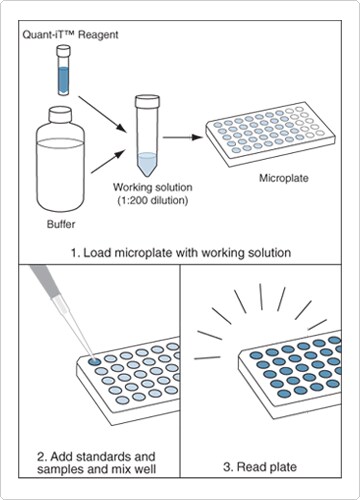
Figure 9.2.1 Microplate assay implementation of DNA, RNA or protein quantitation with the Quant-iT Assay Kits.
Quant-iT Protein Assay Kit
The Quant-iT Protein Assay Kit (Q33210) simplifies protein quantitation without sacrificing sensitivity.![]() This protein assay exhibits a detection range between 0.25 and 5 µg protein, and the response curve is sigmoidal (quasi-linear from 0.5 to 4 µg) with little protein-to-protein difference in signal intensity (Figure 9.2.2). Common contaminants, including salts, solvents, reducing agents (dithiothreitol, 2-mercaptoethanol), amino acids, nucleotides and DNA, are well tolerated in this assay; however, it is not compatible with detergents; slight modifications in the procedure are required for other contaminants. Each Quant-iT Protein Assay Kit contains:
This protein assay exhibits a detection range between 0.25 and 5 µg protein, and the response curve is sigmoidal (quasi-linear from 0.5 to 4 µg) with little protein-to-protein difference in signal intensity (Figure 9.2.2). Common contaminants, including salts, solvents, reducing agents (dithiothreitol, 2-mercaptoethanol), amino acids, nucleotides and DNA, are well tolerated in this assay; however, it is not compatible with detergents; slight modifications in the procedure are required for other contaminants. Each Quant-iT Protein Assay Kit contains:
- Quant-iT protein reagent
- Quant-iT protein buffer
- A set of eight prediluted bovine serum albumin (BSA) standards between 0 and 500 ng/µL
- Easy-to-follow instructions (Quant-iT Protein Assay Kit)
Sufficient reagents are provided to perform 1000 assays, based on a 200 µL assay volume in a 96-well microplate format; this assay can also be adapted for use in cuvettes or 384-well microplates. The fluorescence signal exhibits excitation/emission maxima of 470/570 nm and is stable for three hours at room temperature. The Quant-iT protein reagent is an improved formulation of Molecular Probes NanoOrange reagent, which is described below.
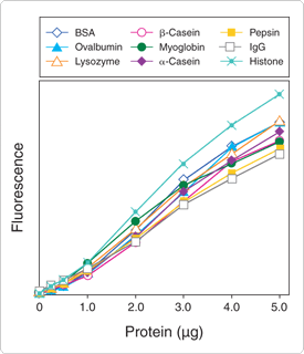
Figure 9.2.2 Low protein-to-protein variation in the Quant-iT protein assay. Solutions of the following proteins were prepared, diluted and assayed with the Quant-iT Protein Assay Kit (Q33210): bovine serum albumin (BSA), chicken-egg ovalbumin, chicken-egg lysozyme, bovine-milk β-casein, equine myoglobin, bovine-milk α-casein, porcine pepsin, mouse immunoglobulin (IgG) and calf-thymus histone. Fluorescence was measured at 485/590 nm and plotted versus the mass of protein sample. Background fluorescence has not been subtracted.
Qubit Protein Assay Kits
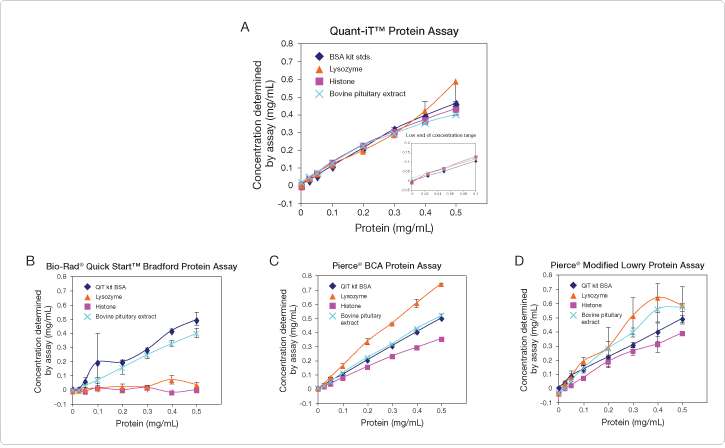
We also offer Qubit Protein Assay Kits specifically designed for use with the Qubit 2.0 fluorometer (100-assay size, Q33211; 500-assay size, Q33212); these kits are also compatible with the original Qubit (1.0) fluorometer. Similar to the Quant-iT Protein Assay Kit described above, the Qubit protein assay compares favorably with other common absorbance- and fluorescence-based assays by providing low protein-to-protein variability, rapid quantitation and accurate results (Figure 9.2.3). The Qubit protein assay is insensitive to many common contaminants, including reducing agents, nucleic acids and free amino acids; however, detergents such as SDS (final concentration >0.01%), Tween 20 and Triton X-100, are not recommended. Each Qubit Protein Assay Kit provides:
- Qubit protein reagent
- Qubit protein buffer
- Three pre-diluted protein standards
- Easy-to-follow instructions (Qubit Protein Assay Kits)
The assay steps are simple: dilute the reagent using the buffer provided, add the sample (any volume between 1 µL and 20 µL is acceptable) and read the concentration using the Qubit 2.0 fluorometer. The assay is performed at room temperature, and the signal is stable for 3 hours. This assay has an optimal detection range of 0.25 to 5 µg, corresponding to initial sample concentrations from 12.5 µg/mL to 5 mg/mL.
We have the Qubit protein assay with three other commonly used assays: the Bio-Rad Quick Start Bradford protein assay, the Pierce BCA protein assay and the Pierce Modified Lowry protein assay. The Bradford method (using Coomassie brilliant blue) exhibited very high protein-to-protein variability (Figure 9.2.3B). Spectrophotometric assays that use BCA (bicinchoninic acid) require carefully timed steps, are not compatible with reducing agents and often yield high estimates of protein, as observed for lysozyme in our study (Figure 9.2.3C). The Lowry method (Figure 9.2.3D) employs a lengthy, multistep procedure and is incompatible with detergents, carbohydrates and reducing agents. The Qubit protein assay showed low protein-to-protein variation, good accuracy and precision and sensitivity down to 0.025 mg/mL in the stock sample (Figure 9.2.3A).
Accurate and sensitive nucleic acid and protein quantitation demands the highest levels of performance, from the analyte-specific assay straight through to the analytical instrument. The Qubit 2.0 fluorometer (Figure 9.2.4) is our second-generation desktop fluorometer designed to work seamlessly with the Qubit DNA, RNA and protein assays (formerly called Quant-iT assays). Together, they form the Qubit Fluorometric Quantitation Platform, an efficient combination of sophisticated, accurate, and highly sensitive fluorescence-based quantitation assays, along with an extremely user-friendly fluorometer. This powerful pairing offers:
- Selective quantitation—more selective and accurate than UV absorbance readings
- High sensitivity—use as little as 1 µL of sample for quantitation
- Intuitive integrated platform—sophisticated quantitation in 5 minutes or less
The Qubit 2.0 fluorometer offers state-of-the art optical components, sophisticated data analysis algorithms and a small bench footprint. It provides the same accuracy provided by the original Qubit (1.0) fluorometer, but with even more speed and less effort than before. The Qubit 2.0 fluorometer has an upgraded, robust design, complete with a color touch screen and intuitive user interface. Navigation buttons for running samples, calibrating standards, and reviewing results are continually accessible at the bottom of the screen, eliminating multiple steps and confusion. And no more scrolling through selections one-by-one using “Up” or “Down” arrows—the Qubit 2.0 fluorometer is equipped with list wheels and scroll bars for clear and simple line selection. Importantly, all functionality has been validated for convenient use with gloves, such as latex and nitrile, commonly worn in a lab setting.
Additionally, the Qubit 2.0 fluorometer includes a new graphing feature that shows the standard curve after calibration and displays up to 10 data points. Sample data is automatically stored in a .csv file format and can be accessed at any time by navigating to the data screen. This stored data file contains all relevant sample information, including time/date stamp, concentration readout, units, and assay type, as well as raw fluorescence data for assay troubleshooting. A built-in dilution calculator allows users to quickly determine sample content, and concentrations determined using this calculator are also stored in the data file. Furthermore, although each Qubit assay function comes pre-programmed with default unit readouts, the Qubit 2.0 fluorometer enables users to change unit output based on their own preference.
For simpler identification and sorting, the data files can be renamed directly using the instrument‘s full alphanumeric keypad. This data can then be transferred to a computer through a USB drive for more thorough analysis—no direct connection to a PC is required at anytime, neither for instrument operation nor data transfer. Likewise, any upgrades can easily be made using a USB drive and firmware downloaded from www.invitrogen.com/qubit. For convenience, every Qubit 2.0 fluorometer comes with a 2 GB USB drive equipped with .pdf files of the complete instrument user manual and all Qubit Assay Kit protocols. We offer the Qubit fluorometer with USB drive separately (Q32866; replacement USB drive, Q32867; replacement international power cord, Q32868) or bundled in a starter kit that includes four different Qubit assay kits as well as a supply of Qubit assay tubes (Q32871). A set of 500 Qubit assay tubes is also available separately (Q32856).
Qubit Assay Kits use advanced Molecular Probes fluorophores that become fluorescent upon binding to DNA, RNA or protein. The selectivity of these interactions ensures more accurate results than can be obtained with UV absorbance readings; Qubit Assay Kits only report the concentration of the molecule of interest, not sample contaminants. Qubit Assay Kits are up to 1000 times as sensitive as UV absorbance readings, and as little as 1 µL of sample is all that's needed for accurate, reliable quantitation. We offer Qubit Assay Kits for a variety of quantitation needs:
- Qubit dsDNA Broad-Range (BR) and High-Sensitivity (HS) Assay Kits—for sequencing samples, genomic DNA samples and routine cloning experiments (Nucleic Acid Quantitation in Solution—Section 8.3)
- Qubit RNA Assay Kits—for microarray experiments, real-time PCR samples and northern blot analysis (Nucleic Acid Quantitation in Solution—Section 8.3)
- Qubit Protein Assay Kits—for protein samples destined for SDS-PAGE, 2D gel electrophoresis and western blot analysis (see above)
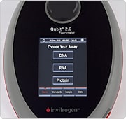
Figure 9.2.4 The Qubit 2.0 fluorometer.
The NanoOrange Protein Quantitation Kit (N6666) provides an ultrasensitive assay for measuring the concentration of proteins in solution.![]() The NanoOrange Protein Quantitation Kit has several important features:
The NanoOrange Protein Quantitation Kit has several important features:
- Ease of use. The NanoOrange assay protocol is much easier to perform than the Lowry method (Figure 9.2.5). Protein samples are simply added to the diluted NanoOrange reagent in a lipid-containing medium, and the mixtures are heated at 95°C for 10 minutes. After cooling the mixtures to room temperature, their fluorescence emissions are measured directly. The interaction of the lipid-coated proteins with the NanoOrange reagent produces a large fluorescence enhancement that can be used to generate a standard curve for protein determination; fluorescence of the reagent in aqueous solutions in the absence of proteins is negligible.
- Sensitivity and effective range. The NanoOrange assay can detect proteins at a final concentration as low as 10 ng/mL when a standard spectrofluorometer or minifluorometer is used. A single protocol is suitable for quantitating protein concentrations between 10 ng/mL and 10 µg/mL—an effective range of three orders of magnitude (Figure 9.2.6).
- Stability. The NanoOrange reagent and its protein complex have high chemical stability. In contrast to the Bradford and BCA assays, readings can be taken for up to six hours after sample preparation with no loss in signal, provided that samples are protected from light.
- Little protein-to-protein variability (Figure 9.2.7). The NanoOrange assay is not only more sensitive, but shows less protein-to-protein variability than Bradford assays.
- Insensitivity to sample contaminants. Unlike the Lowry and BCA assays, the NanoOrange assay is compatible with the presence of reducing agents. Furthermore, the high sensitivity of the assay and stability of the protein–dye complex make it possible to dilute out most potential contaminants, including detergents and salts. The maximum tolerance level for DNA contamination within the NanoOrange assay is 100 ng/mL. Although unusually high concentrations of lipids in the sample can interfere with the NanoOrange assay, this interference can be eliminated by acetone precipitation of the protein, followed by delipidation with diethyl ether.
The NanoOrange protein quantitation reagent, with an excitation/emission maxima of 470/570 nm when bound to proteins, is suitable for use with a variety of instrumentation. Fluorescence is typically measured using instrument settings or filters that provide excitation/emission at ~485/590 nm, which are commonly available for both spectrofluorometers and microplate readers. A spectrofluorometer—either a standard fluorometer or a minifluorometer—offers the greatest effective range and lowest detection limits for this assay. With fluorescence microplate readers, the NanoOrange assay is useful over a somewhat narrower range—from 100 ng/mL to 10 µg/mL in final protein concentration.
The NanoOrange Protein Quantitation Kit (N6666) supplies:
- Concentrated NanoOrange reagent in dimethylsulfoxide (DMSO)
- Concentrated NanoOrange diluent
- Bovine serum albumin (BSA) as a protein reference standard
- Detailed protocols for protein quantitation (NanoOrange Protein Quantitation Kit)
The amount of dye supplied in this kit is sufficient for ~200 assays using a 2 mL assay volume and a standard fluorometer or minifluorometer, or ~2000 assays using a 200 µL assay volume and a fluorescence microplate reader.
The NanoOrange reagent is ideal for quantitating protein samples before gel electrophoresis ![]() and western blot analysis.
and western blot analysis.![]() It has also been used to measure bound versus free protein levels in protein binding assays, and was even able to detect protein trapped in filters during a separation step.
It has also been used to measure bound versus free protein levels in protein binding assays, and was even able to detect protein trapped in filters during a separation step.![]() The NanoOrange reagent is also an optimal reagent for detecting proteins that have been separated by microchip capillary electrophoresis.
The NanoOrange reagent is also an optimal reagent for detecting proteins that have been separated by microchip capillary electrophoresis.![]() A high-throughput assay that may be suitable for clinical samples has been developed for quantitating human serum albumin using a fluorescence microplate reader and using capillary electrophoresis laser-induced fluorescence
A high-throughput assay that may be suitable for clinical samples has been developed for quantitating human serum albumin using a fluorescence microplate reader and using capillary electrophoresis laser-induced fluorescence ![]() (CE-LIF). Additionally, the NanoOrange reagent has been shown to be useful in cell-based assays, including an assay designed to measure total protein content of cell cultures
(CE-LIF). Additionally, the NanoOrange reagent has been shown to be useful in cell-based assays, including an assay designed to measure total protein content of cell cultures ![]() and a rapid method for demonstrating flagellar movement of bacteria.
and a rapid method for demonstrating flagellar movement of bacteria.![]()

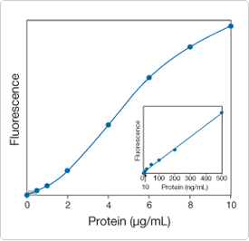
Figure 9.2.6 Quantitative analysis of bovine serum albumin (BSA) using the NanoOrange Protein Quantitation Kit (N6666). Fluorescence measurements were carried out on an SLM SPF-500C fluorometer using excitation/emission wavelengths of 485/590 nm. The inset shows an enlargement of the results obtained (0–500 ng protein per mL) and illustrates the detection limit of ~10 ng/mL.
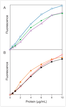
Figure 9.2.7 Quantitative analysis of six different proteins using the NanoOrange Protein Quantitation Kit (N6666): A) bovine serum albumin (BSA, ![]() ), trypsin (
), trypsin (![]() ) and carbonic anhydrase (
) and carbonic anhydrase (![]() ); B) IgG (
); B) IgG (![]() ), streptavidin (
), streptavidin (![]() ) and RNase A (
) and RNase A (![]() ). The y-axis fluorescence intensity scale is the same in both panels, illustrating the minimal protein-to-protein staining variation of the NanoOrange assay. Data were collected using a microplate reader with excitation/emission wavelengths set at 485 ± 20 nm/590 ± 35 nm.
). The y-axis fluorescence intensity scale is the same in both panels, illustrating the minimal protein-to-protein staining variation of the NanoOrange assay. Data were collected using a microplate reader with excitation/emission wavelengths set at 485 ± 20 nm/590 ± 35 nm.
The ATTO-TAG CBQCA reagent (A6222) was originally developed as a chromatographic derivatization reagent for amines ![]() (Reagents for Analysis of Low Molecular Weight Amines—Section 1.8), but this reagent is also useful for quantitating proteins by virtue of its rapid and quantitative reaction with their accessible amines. We have developed the CBQCA Protein Quantitation Kit (C6667, Figure 9.2.8, Figure 9.2.9), which employs the ATTO-TAG CBQCA reagent for rapid and sensitive protein quantitation in solution
(Reagents for Analysis of Low Molecular Weight Amines—Section 1.8), but this reagent is also useful for quantitating proteins by virtue of its rapid and quantitative reaction with their accessible amines. We have developed the CBQCA Protein Quantitation Kit (C6667, Figure 9.2.8, Figure 9.2.9), which employs the ATTO-TAG CBQCA reagent for rapid and sensitive protein quantitation in solution ![]() (A comparison of reagents for detecting and quantitating proteins in solution—Table 9.1). The CBQCA protein quantitation assay functions well in the presence of lipids and detergents,
(A comparison of reagents for detecting and quantitating proteins in solution—Table 9.1). The CBQCA protein quantitation assay functions well in the presence of lipids and detergents,![]() substances that interfere with many other protein determination methods.
substances that interfere with many other protein determination methods.![]() For example, the CBQCA-based assay can be used directly to determine the protein content of lipoprotein samples or lipid–protein mixtures (Figure 9.2.10). The CBQCA assay has been shown to give faster and more sensitive detection of both free amino acids in human plasma
For example, the CBQCA-based assay can be used directly to determine the protein content of lipoprotein samples or lipid–protein mixtures (Figure 9.2.10). The CBQCA assay has been shown to give faster and more sensitive detection of both free amino acids in human plasma ![]() and both low and high molecular weight primary amines in clinical samples from hemodialysis.
and both low and high molecular weight primary amines in clinical samples from hemodialysis.![]() ATTO-TAG CBQCA is more water soluble than either fluorescamine or o-phthaldialdehyde and much more stable in aqueous solution than fluorescamine. Moreover, ATTO-TAG CBQCA provides greater sensitivity for protein quantitation in solution than either fluorescamine or o-phthaldialdehyde (Figure 9.2.11). As little as 10 ng of BSA can be detected in a 100–200 µL assay volume using a fluorescence microplate reader, and the effective range extends up to 150 µg (Figure 9.2.9). Alternatively, the reaction mixtures can be diluted to 1–2 mL for fluorescence measurement in a standard fluorometer or minifluorometer.
ATTO-TAG CBQCA is more water soluble than either fluorescamine or o-phthaldialdehyde and much more stable in aqueous solution than fluorescamine. Moreover, ATTO-TAG CBQCA provides greater sensitivity for protein quantitation in solution than either fluorescamine or o-phthaldialdehyde (Figure 9.2.11). As little as 10 ng of BSA can be detected in a 100–200 µL assay volume using a fluorescence microplate reader, and the effective range extends up to 150 µg (Figure 9.2.9). Alternatively, the reaction mixtures can be diluted to 1–2 mL for fluorescence measurement in a standard fluorometer or minifluorometer.
Each CBQCA Protein Quantitation Kit (C6667) contains:
- ATTO-TAG CBQCA detection reagent
- Potassium cyanide
- Dimethylsulfoxide (DMSO)
- Bovine serum albumin (BSA) protein reference standard
- Detailed protocols for protein quantitation (CBQCA Protein Quantitation Kit)
The CBQCA Protein Quantitation Kit provides sufficient reagents for 300–800 assays using a standard fluorometer, minifluorometer or fluorescence microplate reader.

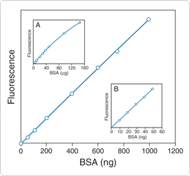
Figure 9.2.9 Detection of bovine serum albumin (BSA) using the CBQCA Protein Quantitation Kit (C6667). The primary plot shows detection of BSA from 50 ng to 1000 ng. Inset A shows that the detection range can extend up to 150 µg. Inset B shows that the lower detection limit can extend down to 10 ng. Each point is the average of four determinations.
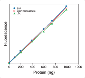
Figure 9.2.10 Quantitation of proteins in a lipid environment using the CBQCA Protein Quantitation Kit (C6667). The protein concentrations of an LDL preparation and a bovine brain homogenate were first determined by the modified Lowry method using BSA as a standard. Assays were then performed using the CBQCA Protein Quantitation Kit on samples containing from 100 ng to 1000 ng protein in 0.1 M sodium borate buffer, pH 9.3, containing 0.1% Triton X-100. Similar results were obtained without the addition of detergent (data not shown). Fluorescence was measured using a fluorescence microplate reader with excitation at 485 ± 10 nm and emission detection at 530 ± 12.5 nm. Each point is the average of three determinations.
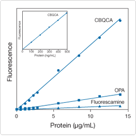
Figure 9.2.11 Comparison of the fluorometric quantitation of bovine serum albumin (BSA) using ATTO-TAG CBQCA (which is supplied in the CBQCA Protein Quantitation Kit, C6667), OPA (P2331MP) or fluorescamine (F2332, F20261). BSA samples were derivatized using large molar excesses of the fluorogenic reagents and were analyzed using a fluorescence microplate reader. Excitation/emission wavelengths were 360/460 nm for OPA and fluorescamine and 485/530 nm for ATTO-TAG CBQCA. The inset shows an enlargement of the results obtained using CBQCA to assay protein concentrations between 0 and 500 ng/mL.
The EZQ Protein Quantitation Kit (R33200) provides a fast and easy high-throughput assay for proteins. Because detergents, reducing agents, urea and tracking dyes do not interfere, this fluorescence-based protein quantitation assay is ideal for determining the protein concentration of samples prior to polyacrylamide gel electrophoresis.![]() This convenient kit can also provide a quick assessment of protein content during protein purification schemes and fractionation procedures.
This convenient kit can also provide a quick assessment of protein content during protein purification schemes and fractionation procedures.
The EZQ assay requires only 1 µL of a sample per spot, and up to 96 samples, including standards, can be assayed in one session. The protein samples are simply spotted onto one of the provided assay papers, fixed with methanol and then stained with the EZQ protein quantitation reagent. This assay paper is then clamped to the specially designed 96-well microplate superstructure for quick analysis in a top- or bottom-reading fluorescence microplate reader. For added versatility, the solid-phase assay format and provided 96-well microplate are also compatible with laser scanners equipped with 450, 473 or 488 nm lasers and with UV illuminators in combination with photographic or CCD cameras for image documentation and analysis. Once the samples are spotted, the assay protocol can be completed in about 1 hour. The protein concentration is determined from a standard curve, and the effective range for the assay is generally 0.05–5 mg/mL or 0.05–5 µg per spot (Figure 9.2.12). Protein-to-protein sensitivity differences in the assay are minimal—the observed coefficient of variation is typically ~16% (Figure 9.2.13).
Each EZQ Protein Quantitation Kit contains:
- EZQ protein quantitation reagent
- EZQ 96-well microplate cassette
- Assay paper
- Ovalbumin, for preparing protein standards
- Detailed protocols for protein quantitation using a variety of fluorescence-detection instruments (EZQ Protein Quantitation Kit)
Sufficient reagent and assay paper are provided for ~2000 protein quantitation assays.
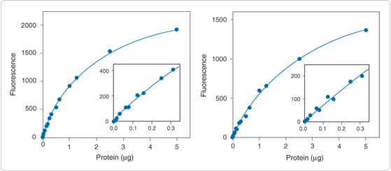
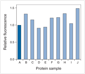
Figure 9.2.13 Protein-to-protein variation in the EZQ protein quantitation assay. Triplicate 1 µg samples of various proteins were assayed using the EZQ Protein Quantitation Kit (R33200) and a fluorescence microplate reader. The mean fluorescence values, after correcting for background fluorescence, are expressed relative to that of ovalbumin. The coefficient of variation is ~16%. The protein samples are: A, ovalbumin; B, bovine serum albumin (BSA); C, myoglobin; D, soybean trypsin inhibitor; E, β-casein; F, carbonic anhydrase; G, transferrin; H, mouse IgG; I, lysozyme; and J, histones.
Most traditional fluorogenic reagents for general protein quantitation in solution detect accessible primary amines. The sensitivity of assays based on these reagents therefore depends on the number of amines available—a function of both the protein's three-dimensional structure and its amino acid composition. For example, horseradish peroxidase (MW ~40,000 daltons), which has only six lysine residues,![]() will be detected less efficiently than egg white avidin (MW ~66,000 daltons), which has 36 lysine residues,
will be detected less efficiently than egg white avidin (MW ~66,000 daltons), which has 36 lysine residues,![]() and bovine serum albumin (MW ~66,000 daltons), which has 59 lysine residues.
and bovine serum albumin (MW ~66,000 daltons), which has 59 lysine residues.![]() However, the assays are generally rapid and easy to conduct, particularly in minifluorometer and fluorescence microplate reader formats.
However, the assays are generally rapid and easy to conduct, particularly in minifluorometer and fluorescence microplate reader formats.
Certain dyes that detect primary aliphatic amines, including ATTO-TAG CBQCA (A6222), fluorescamine (F2332, F20261) and o-phthaldialdehyde (OPA, P2331MP), have been the predominant reagents for fluorometric determination of proteins in solution (A comparison of reagents for detecting and quantitating proteins in solution—Table 9.1). These same reagents, and others such as naphthalene-2,3-dicarboxaldehyde ![]() (NDA, N1138; Reagents for Analysis of Low Molecular Weight Amines—Section 1.8), have frequently been used for amino acid analysis of hydrolyzed proteins.
(NDA, N1138; Reagents for Analysis of Low Molecular Weight Amines—Section 1.8), have frequently been used for amino acid analysis of hydrolyzed proteins.
Fluorescamine
Fluorescamine (F2332, F20261) is intrinsically nonfluorescent but reacts in milliseconds with primary aliphatic amines, including peptides and proteins, to yield a fluorescent derivative ![]() (Figure 9.2.14). This amine-reactive reagent has been shown to be useful for determining protein concentrations of aqueous solutions
(Figure 9.2.14). This amine-reactive reagent has been shown to be useful for determining protein concentrations of aqueous solutions ![]() and for measuring the number of accessible lysine residues in proteins.
and for measuring the number of accessible lysine residues in proteins.![]() Protein quantitation with fluorescamine is particularly well suited to a minifluorometer or fluorescence microplate reader.
Protein quantitation with fluorescamine is particularly well suited to a minifluorometer or fluorescence microplate reader.![]() Fluorescamine can also be used to detect proteins in gels and to analyze low molecular weight amines by TLC, HPLC and capillary electrophoresis.
Fluorescamine can also be used to detect proteins in gels and to analyze low molecular weight amines by TLC, HPLC and capillary electrophoresis.![]()
o-Phthaldialdehyde
The combination of o-phthaldialdehyde (OPA, P2331MP) and 2-mercaptoethanol provides a rapid and simple method of determining protein concentrations in the range of 0.2 µg/mL to 25 µg/mL ![]() (Figure 9.2.15). As compared with fluorescamine, OPA is both more soluble and stable in aqueous buffers and its sensitivity for detection of peptides is reported to be 5–10 times better.
(Figure 9.2.15). As compared with fluorescamine, OPA is both more soluble and stable in aqueous buffers and its sensitivity for detection of peptides is reported to be 5–10 times better.![]() The OPA assay for lysine content is reasonably reliable over a broad range of proteins.
The OPA assay for lysine content is reasonably reliable over a broad range of proteins.![]() OPA can also be used to detect increases in the concentration of free amines that result from protease-catalyzed protein hydrolysis.
OPA can also be used to detect increases in the concentration of free amines that result from protease-catalyzed protein hydrolysis.![]()

Figure 9.2.15 Fluorogenic amine-derivatization reaction of o-phthaldialdehyde (OPA) (P2331MP).
SYPRO Red and SYPRO Orange Protein Gel Stains
An assay has been reported that uses the SYPRO Red protein gel stain (S6653, S6654; Protein Detection on Gels, Blots and Arrays—Section 9.3) for quantitating total protein content of bacterial cells by flow cytometry.![]() This assay provides an accurate measure of planktonic bacterial biomass in marine samples. Fluorescence of the SYPRO Orange protein gel stain (S6650, S6651; Protein Detection on Gels, Blots and Arrays—Section 9.3) has been used to follow isothermal protein denaturation
This assay provides an accurate measure of planktonic bacterial biomass in marine samples. Fluorescence of the SYPRO Orange protein gel stain (S6650, S6651; Protein Detection on Gels, Blots and Arrays—Section 9.3) has been used to follow isothermal protein denaturation ![]() (Monitoring Protein-Folding Processes with Environment-Sensitive Dyes—Note 9.1) and to selectively stain proteins in biofilms prior to two-photon laser-scanning microscopy.
(Monitoring Protein-Folding Processes with Environment-Sensitive Dyes—Note 9.1) and to selectively stain proteins in biofilms prior to two-photon laser-scanning microscopy.![]() SYPRO Red and SYPRO Orange Protein Gel Stains
SYPRO Red and SYPRO Orange Protein Gel Stains
| Cat # | MW | Storage | Soluble | Abs | EC | Em | Solvent | Notes |
|---|---|---|---|---|---|---|---|---|
| A6222 | 305.29 | F,D,L | MeOH | 465 | ND | 560 | MeOH | 1, 2, 3 |
| F2332 | 278.26 | F,D,L | MeCN | 380 | 7800 | 464 | MeCN | 4 |
| F20261 | 278.26 | F,D,L | MeCN | 380 | 8400 | 464 | MeCN | 4, 5 |
| P2331MP | 134.13 | L | EtOH | 334 | 5700 | 455 | pH 9 | 6 |
| ||||||||
For Research Use Only. Not for use in diagnostic procedures.
