Search Thermo Fisher Scientific
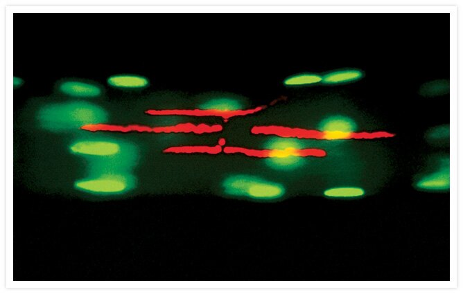
Pseudocolored photomicrograph of the synaptic region of fluorescently labeled living muscle fibers from the lumbricalis muscle of the adult frog Rana pipiens.
Pseudocolored photomicrograph of the synaptic region of fluorescently labeled living muscle fibers from the lumbricalis muscle of the adult frog Rana pipiens. Six hours after isolation of the muscle fibers, acetylcholine receptors were stained with red-fluorescent tetramethylrhodamine a-bungarotoxin (Cat. no. T1175) and myonuclei were stained with the green-fluorescent SYTO® 13 live-cell nucleic acid stain (Cat. no. S7575). Photo contributed by Christian Brösamle, Brain Research Institute, University of Zurich, and Damien Kuffler, Institute of Neurobiology, University of Puerto Rico.

Related Products
Related Images
Developing Drosophila embryo Go ›

Cytoskeleton of a mixed population of granule neurons and glial cells Go ›

Schistosoma mansoni parasite. Go ›

Immunohistochemistry using GFAP Monoclonal Antibody, Mouse, Alexa Fluor® 594 Conjugate Go ›

1% Agarose gel containing 16S and 23S ribosomal RNA (rRNA). SYBR® Green II RNA gel stain. Go ›

Bovine pulmonary artery endothelial cells (BPAEC). MitoTracker® Red CMXRos, SYTOX® Green nucleic acid stain, biotin-XX goat anti–mouse IgG antibody and Cascade Blue® NeutrAvidin biotin-binding protein. Go ›

Live cell imaging with CellLight™ reagents. Go ›

Live cells transduced with Organelle Lights™ or Cellular Lights™ reagents. Go ›

CD335 (NKp46) Antibody (63335182) in RE Go ›

CD223 (LAG-3) Antibody (56223942) in TM Go ›
