Search Thermo Fisher Scientific
Invitrogen
CD20 Monoclonal Antibody (2H7), Brilliant Violet™ 421, eBioscience™
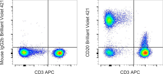
FIGURE: 1 / 1
CD20 Antibody (404-0209-42) in Flow

Product Details
404-0209-42
Species Reactivity
Host/Isotype
Recommended Isotype Control
Class
Type
Clone
Conjugate
Excitation/Emission Max
Form
Concentration
Purification
Storage buffer
Contains
Storage conditions
Shipping conditions
RRID
Product Specific Information
Description: The 2H7 monoclonal antibody reacts with human CD20, a 33-36 kDa transmembrane protein. CD20 is expressed by developing B cells as well as mature B cells but not plasma cells. CD20 has been detected at low levels on a small subset of mature T cells. It is suggested that CD20 plays a role in B-cell activation.
Applications Reported: This 2H7 antibody has been reported for use in flow cytometric analysis.
Applications Tested: This 2H7 antibody has been pre-diluted and tested by flow cytometric analysis of normal human peripheral blood cells. This may be used at 5 µL (0.5 µg) per test. A test is defined as the amount (µg) of antibody that will stain a cell sample in a final volume of 100 µL. Cell number should be determined empirically but can range from 10^5 to 10^8 cells/test.
Brilliant Violet™ 421 (BV421) is a dye that emits at 423 nm and is intended for use on cytometers equipped with a violet (405 nm) laser. Please make sure that your instrument is capable of detecting this fluorochrome.
When using two or more Super Bright, Brilliant Violet™, Brilliant Ultra Violet™, or other polymer dye-conjugated antibodies in a staining panel, it is recommended to use Super Bright Complete Staining Buffer (Product # SB-4401-42) or Brilliant Stain Buffer™ (Product # 00-4409-75) to minimize any non-specific polymer interactions. Please refer to the datasheet for Super Bright Staining Buffer or Brilliant Stain Buffer for more information.
Excitation: 407 nm; Emission: 423 nm; Laser: Violet Laser.
BRILLIANT VIOLET™ is a trademark or registered trademark of Becton, Dickinson and Company or its affiliates, and is used under license. Powered by Sirigen™.
Target Information
CD20 is a non-glycosylated surface phosphoprotein that has a molecular weight range of 33-37 kDa depending on the degree of phosphorylation. CD20 is expressed on mature and most malignant B cells, in a subpopulation of T lymphocytes and follicular dendritic cells. CD20 expression on B cells is synchronous with the expression of surface IgM and it regulates transmembrane calcium conductance, cell cycle progression and B-cell proliferation. CD20 is also associated with lipid rafts, but the intensity of this association depends on extracellular triggering, employing CD20 conformational change, and/or BCR (B cell antigen receptor) aggregation. After the receptor ligation, BCR and CD20 colocalize and then rapidly dissociate before BCR endocytosis, whereas CD20 remains at the cell surface. CD20 serves as a useful target for antibody-mediated therapeutic depletion of B cells, as it is expressed at high levels on most B-cell malignancies, but does not become internalized or shed from the plasma membrane following monoclonal antibody treatment. Diseases associated with CD20 dysfunction include Ms4a1-related common variable immune deficiency.
For Research Use Only. Not for use in diagnostic procedures. Not for resale without express authorization.
How to use the Panel Builder
Watch the video to learn how to use the Invitrogen Flow Cytometry Panel Builder to build your next flow cytometry panel in 5 easy steps.
References (0)
Bioinformatics
Protein Aliases: APY; ATOPY; B-lymphocyte antigen CD20; B-lymphocyte cell-surface antigen B1; B-lymphocyte surface antigen B1; Bp35; CD20; CD20 antigen; CD20 receptor; Fc epsilon receptor I beta chain; Fc Fragment of IgE high affinity I receptor for beta polypeptide; FCER1B; IGEL; IGER; IGHER; LEU16; Leukocyte surface antigen Leu-16; Ly44; Membrane-spanning 4-domains subfamily A member 1; membrane-spanning 4-domains, subfamily A, member 1; MGC3969
Gene Aliases: B1; Bp35; CD20; CVID5; LEU-16; MS4A1; MS4A2; S7
UniProt ID: (Human) P11836
Entrez Gene ID: (Human) 931

Performance Guarantee
If an Invitrogen™ antibody doesn't perform as described on our website or datasheet,we'll replace the product at no cost to you, or provide you with a credit for a future purchase.*
Learn more
We're here to help
Get expert recommendations for common problems or connect directly with an on staff expert for technical assistance related to applications, equipment and general product use.
Contact tech support

