Search Thermo Fisher Scientific
- Order Status
- Quick Order
-
Don't have an account ? Create Account
Search Thermo Fisher Scientific
Beginning in the 1980’s and accelerating in the years since, the use of protein structure information in small molecule drug discovery has significantly reduced the time and cost of bringing molecules to the clinic. Most well-known successes of structure-based drug design efforts (SBDD) are first HIV/AIDS protease inhibitors, which were quickly followed by numerous cancer treatments targeting a range of molecular targets. Despite the growth of biologics modalities, small molecule therapeutics continue to be high value drug assets and comprise the majority of new drug approvals by the Food and Drug Administration (FDA).
Today, the SBDD and Fragment-based drug design (FBDD) approaches have been greatly expanded to new targets and new small molecule modalities owning to the “resolution revolution” in cryogenic electron microscopy (cryo-EM).
In a SBDD approach, the three-dimensional structure of a ligand bound to its target protein is determined with the goal to define and optimize suitable small molecule ligands. Medicinal and computation chemists, together with structural biologists, leverage structures derived from either X-ray crystallography or cryo-EM to improve the drug-target interaction by modifying the shape, size and charge of drug molecules while also identifying areas where ADME profiles can be improved. Structure elucidation of the target protein with and without a small molecule is also critical to inform the mechanism of action (MoA), which is important at all stages of the drug discovery process.

Cryo-EM workflows have dramatically expanded the protein space accessible to structure-based methods, becoming the gold standard for membrane protein structure determination (eg. GPCRs, transporters and channels) and multi-protein complexes. More and more small molecule drugs have been and will be generated due to advances in workflow automaton for high throughput in SBDD using cryo-EM.
Since single-particle cryo-EM has the advantage to reveal high resolution structures of non-crystallizable target proteins, such as membrane proteins and multi-protein complexes, more and more small molecule drugs have been and will be generated due to the advances in workflow automation for high throughput in SBDD using cryo-EM.
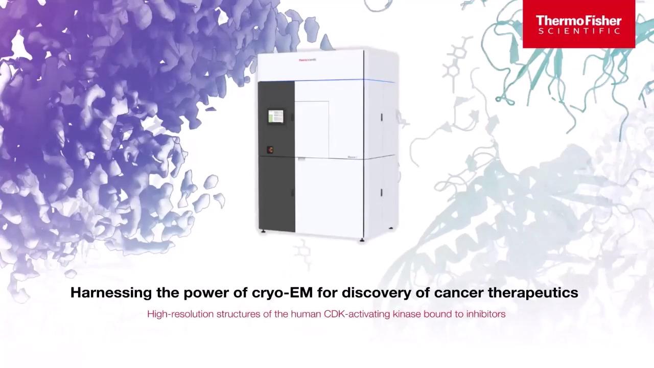
Thermo Fisher Scientific together with Carrick Therapeutics, the Imperial College London and the Institute of Cancer Research successfully developed and published1 a high throughput single particle cryo-EM workflow using the cancer target CDK-activating kinase. With only 1 hour of data acquisition, it was possible to elucidate drug-protein interactions at resolutions of ~3.5–4.5 Å resolution. Extended data acquisition subsequently support structure determination at resolutions up to 1.8 Å atomic resolution.

Workflow utilized for evaluating CDK-targeting compounds. Rapid sample optimization with the Glacios 2 Cryo-TEM is followed by high resolution data collection on a Glacios 2 and/or Krios G4 Cryo-TEM
These structures gave detailed insights into target-inhibitor interactions and display networks of water molecules in the active site of CDK-activating kinase. Due to a proposed mechanism of inhibitor selectivity, this work provides the basis for the rational design of next-generation cancer therapeutics.
Target protein structures at atomic resolution are mandatory for successful lead generation and optimization of small molecule drugs. If the target drug is not known and no preliminary data are available, high-throughput screening of fragment libraries with thousands of small ligands (molecular weight below 500 Da) will accelerate finding suitable ligands with milli to micro molar binding affinities. For an effective FBDD approach, a biophysical method as for example surface plasmon resonance (SPR), isothermal titration calorimetry (ITC) or microscale thermophoresis (MST) pre-screens a large fragment library for determining binding affinities and quantities of ligands bound to the target protein.
Structure elucidation of the ligand-target complexes can today be carried out using cryo-EM. While X-ray crystallography has been the predominant method in FBDD in the past, it displays some clear disadvantages, e.g., protein crystals were dissolved and showed losing diffraction quality in ligand soaking experiments. In addition, challenging drug targets as membrane proteins and protein complexes are typically difficult to crystallization and thus, cryo-EM has become the complementary method for SBDD and FBDD approaches.
Michael Saur et al2., successfully established a FBDD single-particle cryo-EM workflow at Astex Pharmaceuticals. There is a great need in industry to screen fragments for structurally challenging targets such as membrane proteins and protein complexes. Astex Pharmaceuticals created a proof of concept with known multimeric protein targets and screened a small library of fragments using their well-established single-particle cryo-EM workflow.
At an atomic resolution below 2.5 Å, all ligand densities can be clearly identified and show perfect sidechain orientations and chiral configurations. These ligands can be used as building blocks for lead compound generation and optimization and will accelerate drug development of novel small molecules using single-particle Cryo-EM.

Figure from Saur, et. al, under a creative commons license.
In the human genome, only 20% of the target proteins are considered being druggable with bound small molecule drugs. As such, some of the most interesting drug targets, such as transcription factors, RNA-binding proteins, functional multi subunit protein complexes or disease-relevant scaffold proteins, have been excluded by many as targets for small molecule drug development.
An alternative therapeutic approach is to design small molecule ligands which bind to the target protein and recruit it to a E3 ubiquitin ligase leading to ubiquitination and degradation of the target protein via the ubiquitin proteasome system as described by Radhakrishnan et al3. This targeted protein degradation (TPD) approach gives access to a large variety of drug targets in diverse therapeutic areas highlighting it as an emerging market for contemporary and future drug discovery efforts.
The goal in targeted protein degradation is to induce a nearby proximity between the target protein and the E3 ubiquitin ligase. Heterobifunctional protein degraders, also termed proteolysis-targeting chimeras (PROTACs), form a ternary complex between target protein and the ligase inducing a two-way small molecule degrader interaction.

Targeted protein degradation through proximity-inducing compounds. A bivalent molecular degrader forms a ternary complex with an E3 ligase (pink) and a target protein (blue) which leads to attachment of ubiquitin chains (yellow) and subsequent proteasomal degradation. Figure from Radhakrishnan et al, under creative commons license.
In bacteria, target proteins are phosphorylated at arginine residues and degraded via the ClpCP protease system. As a similar therapeutic approach, bacterial PROTACs (BacPROTACs) recruit the target protein to induce TPD. Morreale et al4 used high-resolution single-particle Cryo-EM to reveal the structure of ClpC hexamer in complex with the unfolded BacPROTAC-tethered protein target giving high value structural insights into reprogramming of bacterial ClpCP proteases that could be also confirmed in in vivo experiments using Mycobacteria as a model system. The BacPROTAC induced degradation (BID) technology can be used to degrade attractive protein targets of interest via design and development of novel antibiotics and antimicrobial drugs.

Cryo-EM was utilized to evaluate small-molecule degraders (BacPROTACS). Figures from Morreale et al under creative commons license.

The panel shows a Cryo-EM structure of the activated ClpC hexamer in complex with a BacPROTAC-tethered substrate. Figures from Morreale et al under creative commons license.
A second TPD approach includes molecular glue degraders (MGDs) which bind to a target protein, rearrange it into a neo-substrate being recognized by E3 ubiquitin ligase and being degraded by the UPS. Some degraders have been developed in the past without knowing their mechanism of action (MoA).
Novartis successfully elucidated the structure of the anticancer degrader indisulam bound to DCAF15–DDB1–DDA1–RBM39(RRM2) target-ligase complex using single-particle cryo-EM5. Structural analysis identifies indisulam as a molecular glue degrader binding to DCAF15 ligase and revealing the MoA for protein target degradation. This work explains the high value of cryo-EM in future rational design of novel TPD small molecule degraders.

Structure of the human DDB1-DDA1-DCAF15 E3 ubiquitin ligase bound to RBM39 and Indisulam. Structure recreated from PDB 6SJ7.
Our full cryo-TEM portfolio features state-of-the-art technology with a range of automation features designed to extend accessibility, reduce the need for user intervention, and enable easy organization, viewing, and sharing data.

1. Cushing, VI, Koh, A, Feng, J, et al. High-resolution cryo-electron microscopy of the human CDK activating kinase for structure-based drug design. bioRxiv 2023.04.07.536029. https://doi.org/10.1101/2023.04.07.536029
2. Saur, M, Hartshorn, MJ, Dong, J, Reeks, J, et al. Fragment-based drug discovery using cryo-EM. Drug Discovery Today, 25, 3, (2020), Pages 485-490, ISSN 1359-6446. https://doi.org/10.1016/j.drudis.2019.12.006.
3. Radhakrishnan, S, Hoff, O, Muellner, MK. Current Challenges in Small Molecule Proximity-Inducing Compound Development for Targeted Protein Degradation Using the Ubiquitin Proteasomal System. Molecules 27, 8119 (2022). https://doi.org/10.3390/molecules27238119
4. Morreale, F, Kleine, S, Leodolter, J, et al. BacPROTACs mediate targeted protein degradation in bacteria. Cell 185, 13 (2022). https://doi.org/10.1016/j.cell.2022.05.009
5. Bussiere, DE, Xie, L, Srinivas, H, et al. Structural basis of indisulam-mediated RBM39 recruitment to DCAF15 E3 ligase complex. Nat Chem Biol 16, 15–23 (2020). https://doi.org/10.1038/s41589-019-0411-6
For Research Use Only. Not for use in diagnostic procedures.