Search Thermo Fisher Scientific
- Order Status
- Quick Order
-
Don't have an account ? Create Account
Search Thermo Fisher Scientific

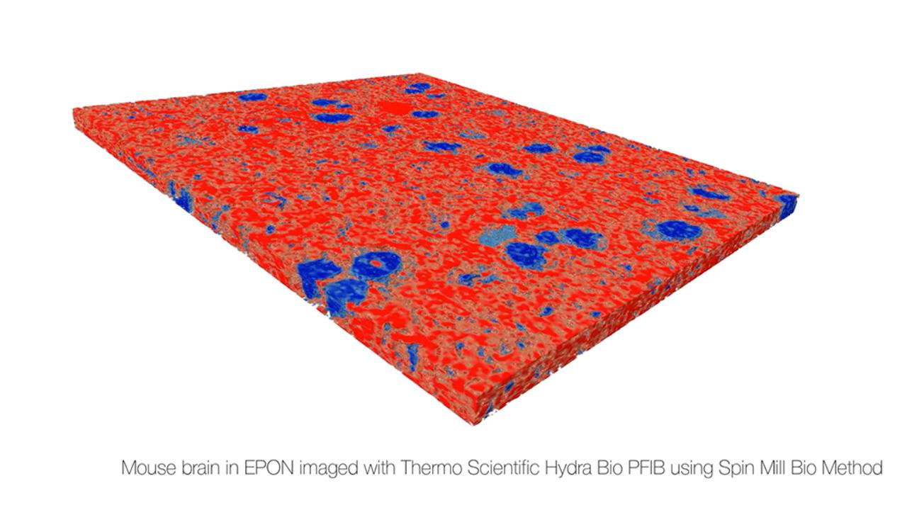
Explore tissues to proteins with the Thermo Scientific Hydra Bio Plasma-FIB. Perform volume imaging at room temperature or in native state with excellent contrast on frozen cells and tissues. Biological samples are fixed in near-native conditions and are stain-free. Bridge the gap between light microscopy and cellular cryo-electron tomography, both in terms of volumes that can be interrogated as well as attainable resolution.

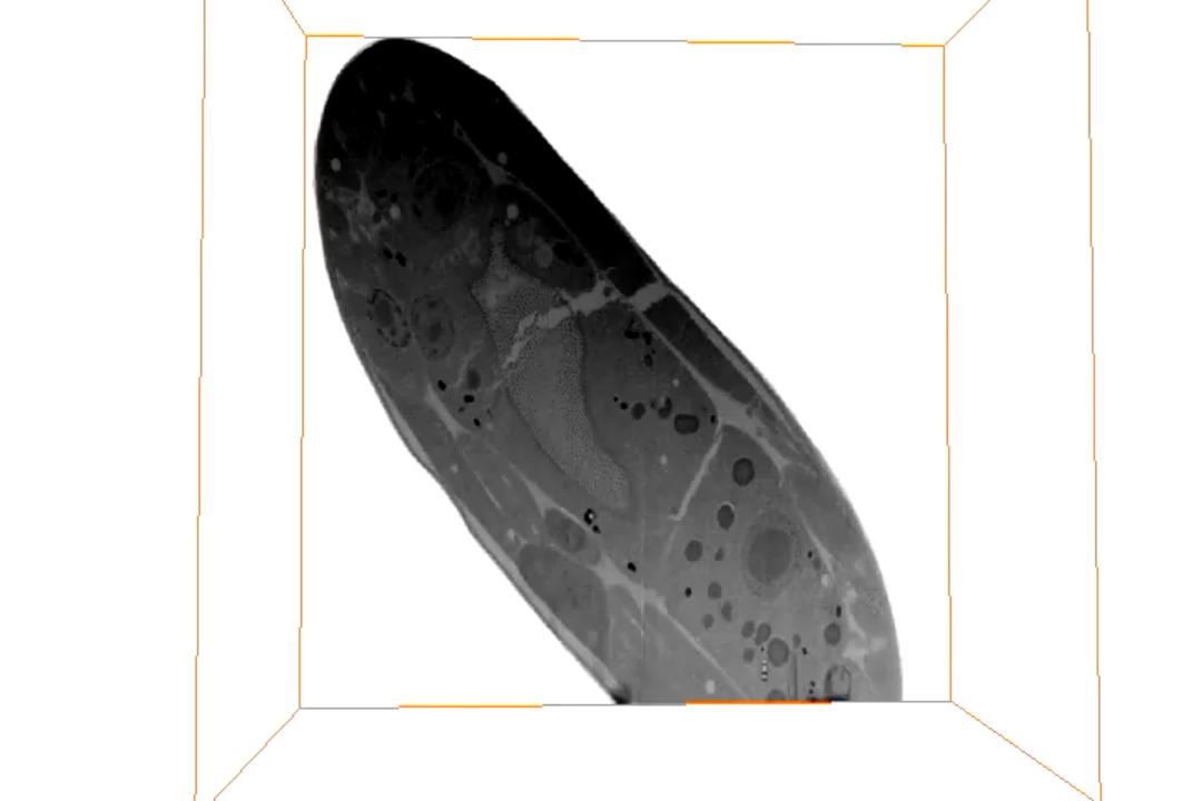
Cryo-electron tomography (cryo-ET) is a technique that allows the study of the 3D structure of cells and tissues at near-native conditions. Sample preparation is a critical step in cryo-ET, which can greatly affect the quality and resolution of the results. Cryo-ET has been applied to many different sample types, from single molecules to protein complexes, viruses, bacteria, cells, to tissue cells and large tissue samples. Samples are either vitrified through plunge-freezing or high pressure freezing (HPF). After the vitrification step, depending on the size of the sample, a thinning step is included in the sample preparation using (plasma) focused ion beam milling to produce cryo-lamellae that are thin enough to be penetrated by electron beams. The Hydra Bio supports automated preparation of lamellae with a thickness of 150 nm or less for the cryo-electron tomography workflow, while avoiding gallium-ion implantation. The guided workflow allows the selection of multiple points of interest and then automatically prepares several lamellae autonomously in unattended runs. The Hydra Bio PFIB prepares lamellae from within large (HPF) volumes with plasma FIB, CLEM localization through integrated light and electron imaging (iFLM Correlative System), and cryo-lift out with Thermo Scientific Easylift Cryo.


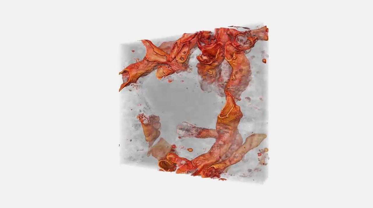
Array tomography can provide rapid localization of cells and their interaction partners in tissues. With the largest imaging field, array tomography is ideal for tissue histology. Ultrastructure analysis of whole animals or whole organs is not needed, so array tomography provides easy navigation for samples that can be stored and re-imaged. Like any Thermo Scientific SEM, the Hydra Bio FIB can be turned into a volume EM microscope with the addition of automated Maps for Array Tomography Software.
For Research Use Only. Not for use in diagnostic procedures.